














Computerized axial transverse scanning was first announced in April 1972 by G.N. Hounsfield.
Computerized axial transverse scanning was first announced in April 1972 by G.N. Hounsfield.
Before we can start begin to review abnormalities in the lungs lets first do a review of normal anatomy.
An owner brings an 8-year-old female, neutered Australian Shepherd with acute right forelimb lameness to your office.
Interventional radiology involves the use of imaging modalities such as fluoroscopy or ultrasonography to gain access to different structures in order to deliver materials for therapeutic purposes.
Q: Please review use of examining low-velocity blood flow in the dog's abdomen.
Blacksburg, Va. -- The Virginia-Maryland Regional College of Veterinary Medicine is offering a new outpatient advanced imaging service for surrounding small-animal veterinary practices starting in June.
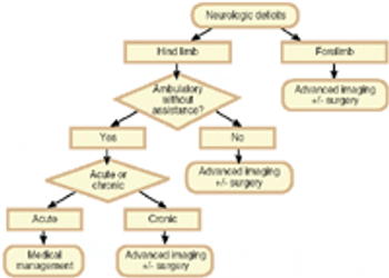
In last month's column we discussed the pathophysiology of intervertebral disk disease (IVDD). Once IVDD is diagnosed, the clinician must again use the history (onset and course of clinical signs) and the physical exam (neurological status) to formulate the therapeutic plan.
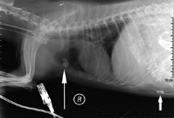
Leakage of intestinal contents and a common contrast medium into a dog's abdominal cavity proved to be a fatal combination.
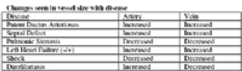
The respiratory system extends from the tip of the nose to the diaphragm.
The soft tissue structures of the joints, back, and foot can be evaluated ultrasonographically yielding important diagnostic information that cannot be obtained radiographically.
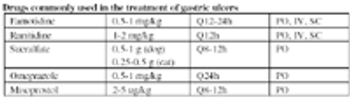
The stomach plays a key initial role in digestion through its mixing actions, and through the secretion of gastric acid and pepsin, which are important for the activation of key digestive enzymes.
Gastrointestinal (GI) cytology offers many advantages to the small animal practitioner in the assessment of patients with gastrointestinal tract disease.
Esophageal diseases, including megaesophagus, can easily sneak up on the unsuspecting clinician if regurgitation, the cardinal sign of esophageal disease, is not considered a differential diagnosis for an animal that presents for what the owner perceives as vomiting.
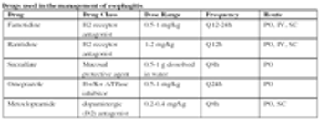
Esophageal foreign bodies and esophagitis have the potential, if not identified and treated, to cause esophageal strictures or megaesophagus, which can be more difficult to treat.
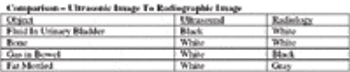
A review of the indications for radiography and ultrasonography.
Primary gastrointestinal (GI) neoplasia is an important differential diagnosis for a dog or cat with vomiting or diarrhea, especially chronic vomiting or diarrhea, anorexia and weight loss, particularly animals that are middle-aged and older.
Diagnostic ultrasonography has, more recently, been applied to the assessment of other less traditional musculoskeletal problems such as evaluation of bone, joints, muscle and nerves.
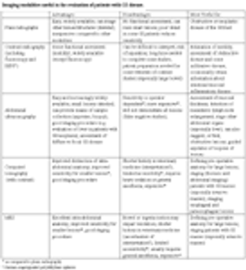
The small animal clinician has a number of imaging options available for the evaluation of dogs and cats with gastrointestinal tract (GI) disease.
Do not buy used "very old" equipment, but equipment that is one generation old is often the best value.
Inflammatory bowel disease is a somewhat loosely defined term that describes chronic gastrointestinal (GI) tract signs in dogs and cats, especially weight loss, vomiting and diarrhea.
Diagnostic imaging is an underutilized resource in herpetological medicine.
Ultrasonography is invaluable in the diagnosis of the cause of colic in horses.
Thoracic ultrasonography yields information about the lung and pleural cavity heretofore unavailable to the veterinarian.
Many of the exotic species we deal with have evolved to mask their illness to avoid predation.
The protein-losing enteropathies (PLE) comprise a collection of intestinal, usually small intestinal, diseases typically associated with weight loss, hypoproteinemia caused by hypoalbuminemia or panhypoproteinemia, and variable signs of weight loss, vomiting and diarrhea.