


Ultrasonography became part of the routine diagnostic method to evaluate pancreatic disorders in small animals at our hospital.
Diagnostics
Latest News
Advertisement
CME Content
Advertisement
For the purpose of this presentation, renal masses are defined as deforming (focal or diffuse) enlargement of the kidney with alteration of the normal renal architecture and/or shape.
In dogs and cats, the most common ultrasonographic feature of gastric tumors is marked thickening of the wall with complete loss of wall layering.
Ultrasonography is part of the routine diagnostic method to evaluate pancreatic disorders in small animals.
The bladder should be thoroughly scanned from left to right in the longitudinal plane and cranial to caudal in the transverse plane.
Radiographs are necessary prior to any US evaluation of the chest.
Overview of Imaging: what you need to know today to better serve your clients tomorrow.
All diseases known fit into a finite number of etiologies: Congenital, Degenerative, Developmental, Inflammatory, Metabolic, Miscellaneous, Neoplastic, Toxic, Traumatic, and Unknown.
Either you control the situation or the situation will control you!
The respiratory system extends from the tip of the nose to the diaphragm.
Disease processes are categorized into Congenital, Degenerative, Developmental Inflammatory, Metabolic, Neoplastic, Traumatic and Miscellaneous processes.
For visualization of soft tissue structures, radiology and ultrasound are complimentary.
The reader tip will help you protect your dental digital radiography sensors.

A recent study sought to establish the incidence of and risk factors for seizures after myelography with iohexol in dogs.
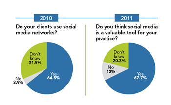
Our study shows that almost 20 percent more veterinary hospitals are using social media compared with last year. Find out how many of your colleagues are posting their way to a better practice.
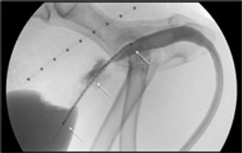
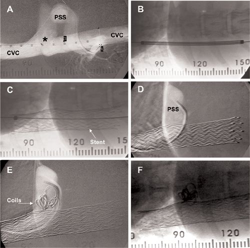
This effective, minimally invasive outpatient procedure can offer immediate relief of stranguria.
Reading radiographs and crime scene investigation require similar skills. Both require identifying a victim, crime, considering the most likely suspects, looking for clues and then building your case or "telling the story". Sticking to these principles will result in a higher conviction rate.
Interpretation of radiographic findings must take patient age and breed into account. Both cats and dogs have typical or age acceptable juvenile and geriatric findings that should not be assumed pathologic. The following is a partial list of age and breed acceptable thoracic findings.
Clipping the hair over the last 2-3 intercostal spaces and extending the area dorsally is important for complete visualization of the liver, particularly in deep chested dog or dogs with small livers.
Clipping the hair over the last 2-3 intercostal spaces and extending the area dorsally is important for complete visualization of the right kidney in the dog.
Clipping the hair and applying alcohol and ultrasound gel is important for maximizing image quality. Ultrasound of the bladder should be performed with the bladder distended; therefore, the patient should not urinate prior to the exam. If the bladder is small and disease is suspected waiting until it fills or filling the bladder with isotonic saline is recommended.
Radiology is a technology driven and technology is rapidly changing. Knowing what is coming soon can alter the diagnostic options we offer clients but keeping up on all the technology can be a full time job. In veterinary radiology we are currently in a digital radiography hotbed.
I'm not telling you something you don't already know when I write that radiographs are not the end all for diagnostic imaging. X-rays were discovered in 1895 and as with any old technology it is typically the least informative. Newer imaging technologies will always provide more information.
GI issues are a common occurrence in the ER and assessment of these cases typically involves imaging, radiographs and/or ultrasound. While obtaining abdominal radiographs is a common and somewhat uncomplicated occurrence, interpretation of abdominal radiographs is anything but.
Abdominal, non-GI issues are frequent in the ER and assessment of these cases typically involves imaging, radiographs and/or ultrasound. While obtaining abdominal radiographs is a common and somewhat uncomplicated occurrence, interpretation of abdominal radiographs is anything but.
Advertisement
Advertisement
Trending on dvm360
1
Tornado (the dog) touches down at the emergency department
2
Every 30 extra minutes under anesthesia raises complication risk in brachycephalic patients, underscoring the importance of peri-anesthetic management
3
Paws and profits: VetEvolve names first chief veterinary officer, NVA appoints two board members, and more updates
4
From the CVO: I’m off the clock
5