Dr. Allison Zwingenberger provides guidelines for identifying foxtails in dogs.
Dr. Allison Zwingenberger provides guidelines for identifying foxtails in dogs.
An owner brings an 8-year-old female, neutered Australian Shepherd with acute right forelimb lameness to your office.

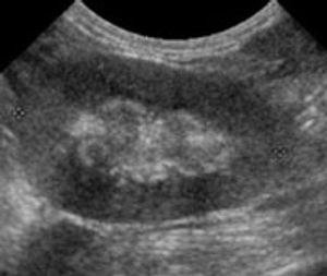
One of the strengths of ultrasound imaging is its ability to resolve very small soft-tissue structures.
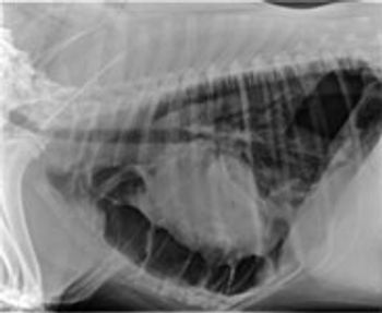
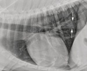
Megaesophagus is a condition in which the esophagus has reduced peristalsis, and has poor tone at rest. The esophagus can have a mild, focal motility problem, or the entire organ may be dilated and functioning poorly.
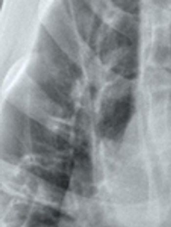
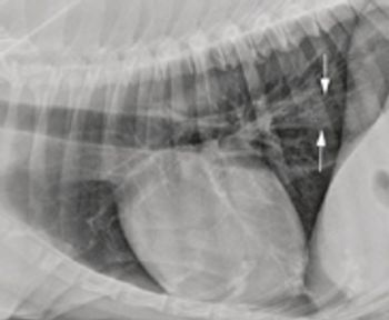
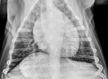
A bronchial pattern on radiographs indicates pathology involving the airways. It can be a subtle pattern to recognize, so let's look at some of the features.

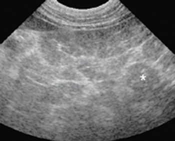
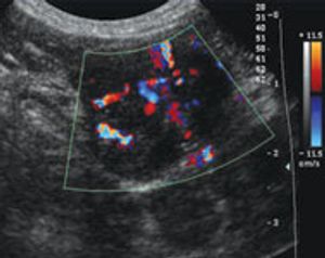
Chronic renal disease is one of the most common ultra-sonographic findings in older cats. If you're doing ultrasounds on cats, you're sure to see signs of chronic renal changes daily.
Thoracic radiographs for metastatic disease are part of every day practice. A diagnosis of pulmonary nodules has an important effect on treatment decisions, and some radiographs are difficult to interpret.
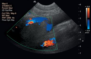
Thrombosis is a complication of many diseases in veterinary medicine.
One of the subtleties of interpreting abdominal radiographs is peritoneal detail.

December 1st 2009
February 1st 2009
August 1st 2008