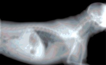
A radiograph can quickly become an expensive and a dangerous waste of time (think of that X-ray exposure!) if its not showing what is needed. Here are some tips to make you a pro.
Michelle Fabiani, DVM, DACVR, Gulf Coast Veterinary SpecialistsDiagnostic Imaging
1111 West Loop SouthHouston, TX 77027

A radiograph can quickly become an expensive and a dangerous waste of time (think of that X-ray exposure!) if its not showing what is needed. Here are some tips to make you a pro.

We often have patients present to us for coughing. Our job is to determine if it is heart disease or pulmonary disease.

Osteochondritis dissecans (osteochondrosis dissecans, OC, OCD) is the most common of the developmental orthopedic diseases and is caused by a sub-condral ossification defect that results in increased thickness of soft articular cartilage, thus decreased nutrient and oxygen availability (from articular fluid) and secondary mechanical failure resulting in a concave bony defect and cartilage flap formation.

Interventional radiology involves the use of imaging modalities such as fluoroscopy or ultrasonography to gain access to different structures in order to deliver materials for therapeutic purposes. The use of interventional techniques in veterinary patients offers a number of advantages compared to more traditional therapies.

Initially it is important to be able to identify radiographic signs of cardiac chamber enlargement. The left atrium on the lateral view when enlarged causes a change in shape of the dorsocadual aspect of the cardiac silhouette.

Sonographic evaluation of the gastrointestinal tract is a routine part of the diagnostic investigation of gastrointestinal disorders. Improved visualization of the GI tract has been achieved due to technologic advances in both ultrasound machines and with the development of higher frequency transducers.

Initially we have to review all the normal structures on a thoracic radiograph before we can begin to discuss pathology. So a review....There are three main normal structures in the lungs: the interstitium, airways, and vessels. The interstitium is the supporting structure of the lungs.

Indications for an esophagram include regurgitation, gagging or retching, dysphagia, cough associated with eating, as well as the presence of a mediastinal, cervical, or thoracic mass. The pertinent anatomy to remember is that in the cat the caudal 1/3 of the esophagus is smooth muscle.

Before pathology can be discussed, the normal appearance of the liver, biliary system, and pancreas will be reviewed. Determination of liver size via US is not accurate and is best done on radiographs. Ultrasound is best performed with the animal in dorsal recumbency (on their back) and the area must be clipped free of hair.

Let's begin with the upper urinary tract – the kidneys and ureters. Knowing normal anatomy is of course initially necessary to perform an adequate ultrasound examination. You should always scan in two planes (sagittal and transverse). The right kidney is harder to visualize as it is located at the level of T13 and is located in the caudate fossa of the liver.

With conventional film-screens, 49% of X-rays pass through the cassette without interaction at all; 50% interact with the phosphor layer of the screen.

The purpose of this talk is to discuss what MR can and cannot image, review the basic neurologic diseases imaged with MR, and ultimately know when to recommend MRI to your patients.

When performing a complete cardiac evaluation, a minimum of two views are necessary: lateral and either VD or DV.

Indications for an esophagram include regurgitation, gagging or retching, dysphagia, cough associated with eating, as well as the presence of a mediastinal, cervical, or thoracic mass.

Computerized axial transverse scanning was first announced in April 1972 by G.N. Hounsfield.

Before we can start begin to review abnormalities in the lungs lets first do a review of normal anatomy.

Interventional radiology involves the use of imaging modalities such as fluoroscopy or ultrasonography to gain access to different structures in order to deliver materials for therapeutic purposes.

Published: August 1st 2009 | Updated:

Published: May 1st 2011 | Updated:

Published: August 1st 2009 | Updated:

Published: August 1st 2009 | Updated:

Published: August 1st 2009 | Updated:

Published: August 1st 2009 | Updated: