


To make cleanup after dental examinations quicker and easier, wrap a clear plastic trash can liner around the head of your dental radiography unit before beginning.
Diagnostics
Latest News
Advertisement
CME Content
Advertisement
Many human and veterinary hospitals are switching to digital radiography systems, so if you are still using film, you may be able to easily acquire their obsolete cassettes and film to supplement your practice's inventory, as I did.
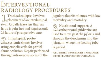
East Lansing, Mich. - The introduction of minimally invasive procedures such as interventional radiology offers the same benefits to animals that similar procedures have made possible in human medicine, according to one practitioner.
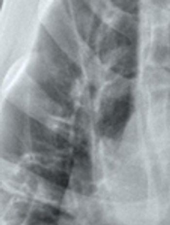
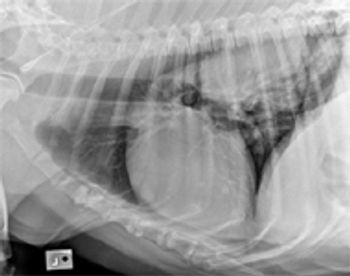
A bronchial pattern on radiographs indicates pathology involving the airways. It can be a subtle pattern to recognize, so let's look at some of the features.
Santa Clara, Calif. - Eklin Medical Systems completed its acquisition of Ultrasource. The deal was announced in late May. Ultrasource is the U.S. veterinary distributor for Philips Medical Systems' ultrasound line, which includes Sonoscape and SIUI.
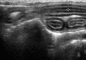
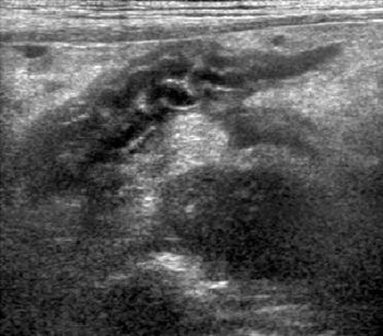
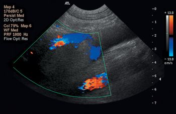
Ultrasound of the gastrointestinal (GI) tract has become an increasingly popular and useful diagnostic procedure for evaluation of gastric and intestinal disease.
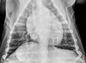
Thoracic radiographs should initially be evaluated for positioning, technique (exposure factors), and the presence of any artifacts that might affect interpretation.
Pancreatitis is a common consideration in dogs and in an increasing number of cats presented for vomiting, anorexia, lethargy, or abdominal pain.
Radiographic interpretation of pulmonary disease is a critical part of veterinary diagnostics, but can be one of the more intimidating areas of radiographic evaluation.
Evaluation of the mediastinum and pleural space is difficult on physical examination, due to the relative inaccessibility of these anatomical areas.
Failure to read all of the radiograph can result in serious oversights.
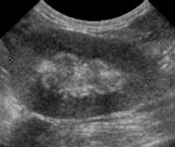
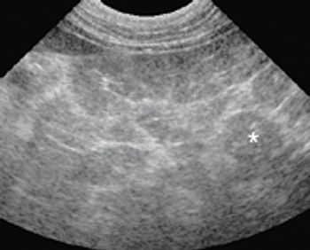
Chronic renal disease is one of the most common ultra-sonographic findings in older cats. If you're doing ultrasounds on cats, you're sure to see signs of chronic renal changes daily.

The measure of a machine isn't just what shows up on a cost-benefit analysis.
College Station, Texas- 3/31/08 - The Texas A&M University Board of Regents has approved the College of Veterinary Medicine & Biomedical Sciences to begin construction on a veterinary imaging and cancer treatment center.
Thoracic radiographs for metastatic disease are part of every day practice. A diagnosis of pulmonary nodules has an important effect on treatment decisions, and some radiographs are difficult to interpret.
In this prospective study from a referral practice, the limbs of 10 healthy dogs were evaluated by using a thermographic imaging protocol to determine normal cutaneous thermographic patterns (a color map that indicates the skin temperature distribution) and evaluate the effect of hair clipping.
In this retrospective study of 100 cases from a veterinary college, preoperative abdominal ultrasonography and exploratory laparotomy findings were reviewed to determine agreement and discrepancy rates and identify lesions likely to be missed by ultrasonography.
Thrombosis is a complication of many diseases in veterinary medicine.
One of the subtleties of interpreting abdominal radiographs is peritoneal detail.
2008 Editorial Calendars
List of Ownership Issues stories in January through December of Veterinary Economics.
Dr. Niemiec illustrates how to obtain dental radiographs using the correct bisecting angle.
Dr. Niemiec demonstrates the right positioning for obtaining dental radiographs of the maxillary canines.
Dr. Niemiec demonstrates how to avoid errors of vertical angulation when taking dental radiographs.
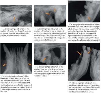
Interpreting dental radiographs is quite similar to interpreting standard radiographs except dental pathologies and radiographic changes may be subtle and some pathologies are unique to the oral cavity. Also, several normal anatomical structures may mimic pathologic changes.
Advertisement
Advertisement
Trending on dvm360
1
Tornado (the dog) touches down at the emergency department
2
Every 30 extra minutes under anesthesia raises complication risk in brachycephalic patients, underscoring the importance of peri-anesthetic management
3
Paws and profits: VetEvolve names first chief veterinary officer, NVA appoints two board members, and more updates
4
From the CVO: I’m off the clock
5