


Incorrect answer for Image Quiz: Cytology-A dyspneic calico with a runny nose
Diagnostics
Latest News
Advertisement
CME Content
Advertisement

A useful protocol for identifying orthopedic lesions.
The best diagnostic plan for feline odonotoclastic resorptive lesions.
Find out how to predict gallbladder rupture in dogs.
PDF ONLY. DO NOT LINK.
PDF ONLY. DO NOT LINK.
WVC clinical speaker discusses how to interpret radiographs quickly and accurately in critical patients.
Chronic vomiting is a common presenting complaint from owners of dogs evaluated at both primary hospitals and referral institutions.
When medical management is no longer adequate, patients with tracheal collapse need more aggressive surgical intervention.

Quiz: Holiday myths debunked (6-Fact)
How to evaluate overexposed areas of a radiograph.
The use of computed tomography (CT), though not readily available at every institution, is becoming a more widespread modality for use in the small animal patient. Computed tomography affords a rapid evaluation of skeletal images with a small slice thickness that can be as small as 0.625.
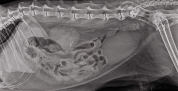
Abdominal radiographs are a rapid, readily available method to give an overview of the abdomen. Though most people believe ultrasound is the new modality of choice for abdominal evaluation, the limitations of ultrasound not being able to penetrate gas as well as the technical ability and time to acquire images still make abdominal radiographs a great first modality in the patient with acute abdominal pain.
The explosion of magnetic resonance imaging (MRI) has revolutionized neuroimaging. However, other modalities still are valuable when looking at the central nervous system. These include ultrasound and computed tomography (CT). Radiographs have limited usefulness due to the superimposition of structures associated with the cranial vault and vertebral column.
The use of radiography to examine the abdomen is full of complications. Radiographs are very good at determining the difference between bone and gas, but soft tissue and fluid are the same opacity. When dealing with intra-abdominal lesions, the main goal is to differentiate one soft tissue mass from a normal soft tissue structure from abdominal fluid. Ultrasound uses high frequency sound waves to accomplish what radiographs cannot.
Ultrasound examinations are becoming routine standard of care in veterinary medicine. Due to the non-invasive nature of the modality, the general affordability of the hardware and the growing amount of continuing education to train general practitioners and veterinary technicians on the technical aspects of scanning, ultrasound is becoming the modality of choice, especially when examining the abdomen.
Diagnosing disease in reptiles can be a challenge for even the most experienced veterinarian. The clinical signs exhibited by these ectotherms are often subtle and physical findings are seldom pathognomonic.
Recognizing that thoracic radiography is the first diagnostic imaging step for dogs and cats with thoracic disease, there is a lot more to thoracic imaging than radiographs. In specific circumstances additional modalities which may be considered include ultrasound, computed tomography (CT), fluoroscopy and nuclear scintigraphy.
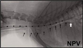
The evaluation of the musculoskeletal system is difficult due to the numerous soft tissues as well as the bone structures involved. Rapid assessment of the bone structure is routinely performed using radiographs; however, the subtlety of disease and joint compared to bone pathology can be confusing.
Diagnostic imaging has seen a huge technology shift in the last 10 years. Modalities that were not accessible to the small animal patient, such as magnetic resonance imaging, are now considered the modality of choice for neurologic examinations. This technology shift has caused a lot of confusion as well as questions about what modalities are used for which diseases and why.
Interventional radiology helped relieve this cat's ureteral obstruction by uroliths.

Could this common fracture eventually be a thing of the past?
An imaging pointer for staging lung cancer.
Advertisement
Advertisement
Trending on dvm360
1
Tornado (the dog) touches down at the emergency department
2
Every 30 extra minutes under anesthesia raises complication risk in brachycephalic patients, underscoring the importance of peri-anesthetic management
3
Paws and profits: VetEvolve names first chief veterinary officer, NVA appoints two board members, and more updates
4
From the CVO: I’m off the clock
5