


Dr. Ryan King provides a brief overview of which cases are best suited for CT vs. MRI.From the CVC in San Diego, veterinary radiologist Dr. Ryan King provides a brief overview of which cases are best suited for computed tomography (CT) vs. magnetic resonance imaging (MRI).
Diagnostics
Latest News
Advertisement
CME Content
Advertisement

Dr. Ryan King outlines which forms of imaging are most helpful in certain clinical cases.

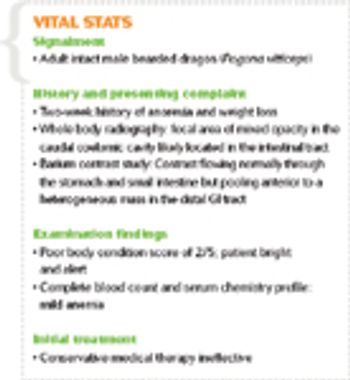
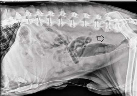
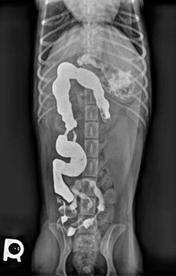
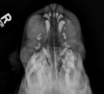
A contrast enema study helped clinicians detect an intestinal stricture in this exotic pet. See how this diagnostic step can help your reptile patients with similar signs.
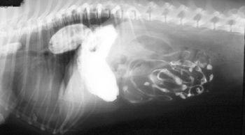
Be sure to look a little deeper when using this technique during your search for a ruptured bladder to prevent missing the diagnosis.
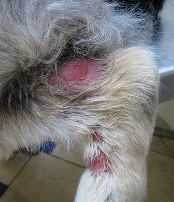
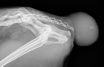
What lies beneath this little dog's bite wounds?
Check out the stories, photos and floor plans of these 10 Merit Award-winning veterinary hospitals.
Client handout: Answers to your questions about pemphigus foliaceus in dogs and cats
Pick the right diagnostic step in this senior dog.
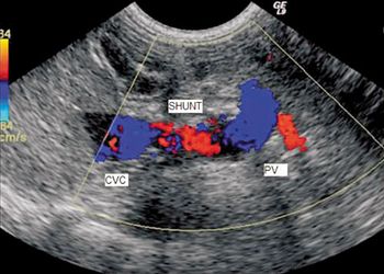
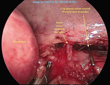
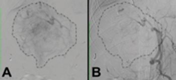
When you have a patient with a hepatic vascular abnormality, how do you confirm it?
Add a little contrast to help you find your diagnosis.
Examine the imaging results to help solve this pug's problem.
Dr. Anthony Pease discusses one aspect of digital systems that you don't have to worry about.

This on-demand course was written to provide information in a fun, easy, and memorable way that can be used to help hone your radiation safety skills; answer questions you may have; give you information you can use to decrease the amount of radiation you, as a radiation worker are exposed to; and to help you prepare for national, state, or local exams if you have those ahead. As people who spend a lot of time leaded, we also included radiation apparel comparisons and our recommendations. (2 CE credits)
In this on-demand CE course, you will become familiar with aspects of ultrasonography that will help veterinarians and technicians prepare patients for ultrasound exams, provide the basic knowledge needed for doing ultrasound exams, and learn terminology associated with ultrasonography. It is not meant to be a replacement for adequate and extensive hands-on training in doing ultrasounds. The course is written by Fern Delaney, a registered diagnostic medical sonographer who specializes in performing veterinary ultrasonography and training other professionals to do so. (2 CE credits)
Can you discover the cause?
A novel veterinary treatment offers hope for these often hopeless cases.
A minimally invasive option to treat this common occurrence in pets.
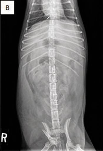
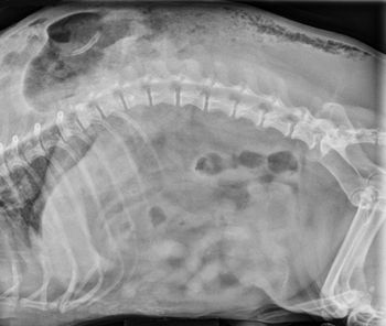
Look at the radiographs and determine the most likely diagnosis.

Make rounds with these veterinary specialists and residents for a complete picture of this neoplasia in dogs.
Advertisement
Advertisement
Trending on dvm360
1
Tornado (the dog) touches down at the emergency department
2
Every 30 extra minutes under anesthesia raises complication risk in brachycephalic patients, underscoring the importance of peri-anesthetic management
3
Paws and profits: VetEvolve names first chief veterinary officer, NVA appoints two board members, and more updates
4
From the CVO: I’m off the clock
5