


Front-desk quiz: Test your medical knowledge answer 1D
Diagnostics
Latest News
Advertisement
CME Content
Advertisement

How to help detect peritoneal disease on radiographs.
To help organize the markers used with radiographs, we purchased a plastic trading-card protector.
Abdominal ultrasound has taken on a "larger-than-life" position in diagnostic imaging in veterinary medicine for several reasons. First and foremost is that ultrasound is a non-invasive technique that can be used by the small animal practitioner for imaging the peritoneum, parenchyma of the abdominal organs and retroperitoneum.
Aside from ultrasound, a digital radiography suite is going to be the most expensive upgrade for the private practitioner in the realm of diagnostic imaging. However, this expense will be well worth the investment from a diagnostic imaging standpoint. After more than a century of film and film screen imaging, the backdrop of diagnostic radiology has changed from hanging films on view boxes to LCD monitors.
This brief overview of abdominal ultrasound is meant to be just that – an overview. If you are serious about ultrasound, you will take it upon yourself to review on the ultrasound references in physics such as that by Kremkau.
Obvious thoracic disease need not be discussed here. Fulminating heart failure, pneumonia, advanced metastatic lung disease, severe pneumothorax and pleural effusion are reliably diagnosed radiographically. What I would like to share with you are the less obvious radiographic manifestations of acute cardio-respiratory disease and my approach to diagnosis. And remember, in the final assessment much can be learned from the presenting clinical signs. Does the patient have a cardiac murmur or history of heart disease? Was there an observed or suspected trauma?
Much can be learned about the gastrointestinal tact (GIT) by careful evaluation of survey abdominal radiographs. Survey radiographs of the abdomen in cases of GIT disease should always include the stomach, liver, and diaphragm. Stomach position, size, shape and contents are evaluated. The small intestine is assessed for position and distribution within the abdominal cavity, diameter, and luminal contents.
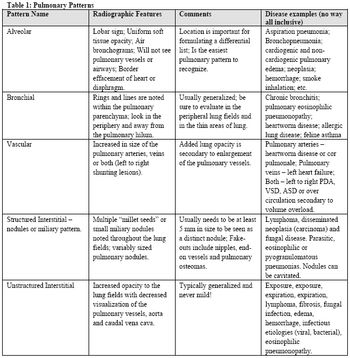
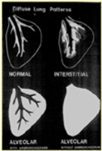
Pulmonary patterns have the bane of radiology since the beginning of time (1896 when x-rays were discovered). Pulmonary pattern recognition is the most difficult concept to teach and the most difficult and frustrating to learn, yet, the pattern itself is only part of the puzzle.
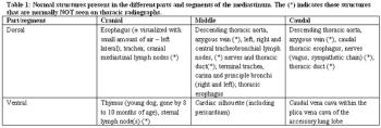
Mediastinal abnormalities, including cardiac disease, are common causes of clinical signs related to the thorax. By definition, the mediastinum is the midline potential space formed between the two pleural cavities and includes the medial portions of the right and left parietal pleura (also called the mediastinal pleural) and the space formed between these serosal membranes.
Thoracic radiography is still the most common first line assessment for diseases and conditions of the thorax. With the advent of digital radiography, a new interest in diagnostic radiology has emerged. However, even though certain artifacts are not an issue (e.g., processing, exposure), problems with inadequately positioned patients still exist.
The normal thorax is well suited to radiographic evaluation because there is marked inherent contrast between the air-filled, fluid-filled, soft tissue, and bony structures that comprise the thoracic viscera and thoracic wall. As has been stated before, at least 2 orthogonal views of the thorax are required for complete and accurate interpretation. For routine evaluation of the thorax, either a right or left lateral projection, and a dorsoventral or ventrodorsal projection of the thorax are required.
Pulmonary patterns have the bane of radiology since the beginning (1896 that is). The most difficult concept to teach and the most difficult to learn, yet, the pattern itself is only part of the puzzle. The recognition that the disease is actual within the pulmonary parenchyma and not in the pleural space, extrathoracic structures or the mediastinum is the first step.
In this session we will review thoracic radiology and echocardiography with an emphasis on normal and abnormal anatomic features.
Radiographic evaluation has fast become a common facet of veterinary dentistry and only practices that utilize dental radiography can practice quality dentistry. Interpretation of radiographic changes that occur in the tooth and surrounding bone take many forms.
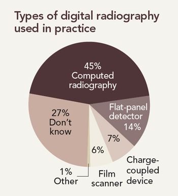
A new study finds that most veterinarians still use film-based radiography, but digital radiography is up and coming.
Aside from ultrasound, a digital radiography suite is going to be the most expensive upgrade for the private practitioner in the realm of diagnostic imaging. However, this expense will be well worth the investment from a diagnostic imaging standpoint. After more than a century of film and film screen imaging, the backdrop of diagnostic radiology has changed from hanging films on view boxes to LCD monitors.
Incorrect answer for Image Quiz: Dermatology-A bulldog with erythematous plaque
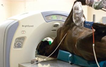
CT scanning has many uses in equine medicine and surgery.
Telemedicine offers profit and high-quality care to clients and practitioners.
The inherent poor contrast within the abdomen and the fact that soft tissue and fluid can not be differentiated radiographically means that contrast media are required for assessment of luminal surfaces, and therefore wall thicknesses of the gastrointestinal tract.
Digital radiography is the fastest growing imaging modality in medicine. It is replacing conventional analog imaging in practices across the United States. Eventually all veterinary practices will utilize this modality.
The thorax is traditionally examined by a compartment approach-6 basic compartments or "spaces" plus the thoracic wall should be considered during the radiographic examination. The compartments include the mediastinum, the pleural space and four pulmonary divisions-bronchial, vascular, interstitial and alveolar.
Computed tomography is becoming more and more readily available to general practices, either as a local referral practice or in-house in larger practices. Typically this procedure is performed only on the most complex cases and only under general anesthesia.
Survey radiography is commonly used to image the urinary tract and provides information on size, shape, opacity, location and, margination of urinary organs. This modality is rapid and cost effective for screening animals with suspected urinary tract disease.
Advertisement
Advertisement
Trending on dvm360
1
Tornado (the dog) touches down at the emergency department
2
Every 30 extra minutes under anesthesia raises complication risk in brachycephalic patients, underscoring the importance of peri-anesthetic management
3
Paws and profits: VetEvolve names first chief veterinary officer, NVA appoints two board members, and more updates
4
From the CVO: I’m off the clock
5