


The patient is coughing with no other signs, but now there is a murmur. What to do? I take radiographs and hope that they guide me for the next test.
Diagnostics
Latest News
Advertisement
CME Content
Advertisement
Radiographic assessment of the heart and pulmonary vessels is challenging regardless of the species. This is due to numerous factors including variation between species and breeds, exposure factors, effects of the cardiac and respiratory cycles, radiographic positioning and quality of x-ray equipment.
Computed tomography is becoming more and more readily available to general practices, either as a local referral practice or in-house in larger practices.
Diagnosis of emergency-critical care conditions requires the same attention to good radiographic technique as routine conditions. Otherwise serious errors can result which may seriously affect outcome.
This session will discuss the relative advantages and disadvantages of digital radiography. This modality can save time, money and frustration, but comes with an initial up-front financial cost.
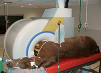
During the last few years, magnetic resonance imaging and computed tomography have made significant improvements in assessing equine tissue damage and diagnosing disease.
A 2-year-old intact male Boston terrier was evaluated because of a recent onset of gagging and vomiting.
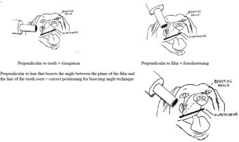
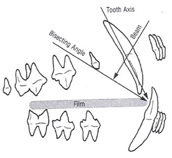
A good clinical oral examination is one of the most important tools we have for diagnosing many dental and oral problems. However, much of the pathology in the oral cavity involves the roots and supportive bone of the teeth instead of their visible crowns.
Radiographs should be made whenever clinical examination indicates that there might be subgingival pathology that could be identified or better characterized by evaluating the hidden hard tissues.

At first glance, Red Bank Veterinary Hospital in Tinton Falls, N.J., seems like an intimidating behemoth of a building. Look inside, though, and you'll find an efficient layout and a helpful staff ready to provide top-notch care for pets.
Most incidents involve dogs that accidentally ingest drugs left out in the home.
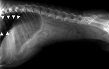
Although aspiration pneumonia is a common clinical diagnosis in dogs, studies on the causes, diagnostic findings, and outcome of affected dogs are sparse.
A new development in the diagnosis of upper airway disease
Dr. Ravinder Dhaliwahl says most veterinary oncologists prefer three thoracic radiographic views.
This lecture is a case-based presentation of subtleties noted on radiographs that indicate a problem occurring on some other area of anatomy not directly visible on the radiographs.
The lecture is a review of thoracic radiographic interpretation in small animals.

This on-demand CE course includes the Dr. Amy S. Lang's top 10 tips for professional and good quality radiographs. (1 CE credit)
If you find elbow radiography challenging or you want to pick up some little known tips for better results, this on-demand CE course is for you. (1 CE credit)
This on-demand CE course contains ideas to help improve the quality of abdominal radiographs. (1 CE credit)
This on-demand CE course contains tips and information that can be used to improve the quality of thorax radiographs. (1 CE credit)
This on-demand CE course on digital radiography covers digital terminology, equipment, which features you might want, and more. (1 CE credit)
This on-demand CE course covers hip positioning, technique, views, and tips beyond what is covered by traditional information sources. (1 CE credit)

Participation in this CE offering is available to members of the National Association of Veterinary Technicians in America. The applicable article is available in the September 2008 issue of The NAVTA Journal. (1 CE credit)
With conventional film-screens, 49% of X-rays pass through the cassette without interaction at all; 50% interact with the phosphor layer of the screen.
Veterinary dentistry is emerging as one of the fastest growing "new" disciplines of veterinary patient care.
Advertisement
Advertisement
Trending on dvm360
1
Tornado (the dog) touches down at the emergency department
2
Every 30 extra minutes under anesthesia raises complication risk in brachycephalic patients, underscoring the importance of peri-anesthetic management
3
Paws and profits: VetEvolve names first chief veterinary officer, NVA appoints two board members, and more updates
4
From the CVO: I’m off the clock
5