


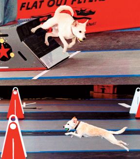
Dr. Wendy Baltzer provides examples of appropriate warm-up and cool-down exercises for agility dogs.
Orthopedics
Latest News
Advertisement
CME Content
Advertisement
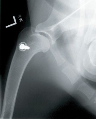
Raleigh, N.C. - A veterinary team at North Carolina State University recently performed a unique total knee replacement surgery on a cat.
In this photo gallery, veterinary surgeon Scott Palmer demonstrates how he straightened and lengthened a disfigured leg of a Great Dane puppy by using a novel surgical technique.
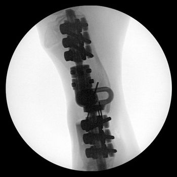
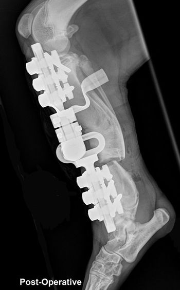
Fort Collins, Colo. - To save a dog's leg, a veterinary orthopedic surgeon tried something radical.
This is a question in "BizQuiz: Is stationary or mobile equine veterinary practice right for you?"
This is a question in "BizQuiz: Is stationary or mobile equine veterinary practice right for you?"
This is a question in "BizQuiz: Is stationary or mobile equine veterinary practice right for you?"
This is a question in "BizQuiz: Is stationary or mobile equine veterinary practice right for you?"
Image Quiz: Dermatology-The case of the blind Akita
The search for antibiotics began in the late 1800s, with the growing acceptance of the germ theory of disease, a theory that linked bacteria and other microorganisms to the causation of a variety of ailments.
Lameness and pain caused by osteoarthritis (OA) is one of the most common presenting complaints in small animal practice.
Juvenile pubic symphysiodesis: a simple, affordable surgical solution to canine hip dysplasia.
Juvenile pubic symphysiodesis (JPS) is a minimally invasive, affordable, prophylactic procedure performed in immature dogs at risk for development of arthritis associated CHD.
Feline osteoarthritis (OA) is a growing problem in our veterinary patients. We are discovering that it has been around and under diagnosed for years.
Panosteitis is an acquired self-limiting condition of undetermined cause that affects the diaphyseal and metaphyseal regions of the long bones of young, large breed dogs.
Cranial cruciate ligament rupture is a common cause of hindlimb lameness in dogs and is seen in cats as well.
CCL injury is one of the most common injuries encountered by veterinarians, and is by far the most common cause of lameness of the sti?e joint.
At birth the hips are normal. The femoral head and neck are cartilaginous and begin forming bone by endochondral ossification.
Coxofemoral luxation is the most commonly luxated joint in dogs, accounting for 90% of all luxations. It is usually the result of trauma or severe hip dysplasia with 78% being craniodorsally luxated.
At birth most hips are normal. The femoral head and neck are cartilaginous and begin forming bone by endochondral ossification.
Physical rehabilitation is becoming a common place therapy in veterinary medicine. Several benefits have been proven and continue to be elucidated.
Incorrect answer for Image Quiz: Dermatology-A dog with a suddenly crusty, itchy muzzle
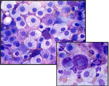
Correct answer for Image Quiz: Cytology-A subcutaneous mass in a senior dog

Answers to frequently asked questions you need to know.
Advertisement
Advertisement
Trending on dvm360
1
Every 30 extra minutes under anesthesia raises complication risk in brachycephalic patients, underscoring the importance of peri-anesthetic management
2
From the CVO: I’m off the clock
3
Tornado (the dog) touches down at the emergency department
4
Paws and profits: VetEvolve names first chief veterinary officer, NVA appoints two board members, and more updates
5