R.D. Montgomery, DVM, MS, DACVS
Articles by R.D. Montgomery, DVM, MS, DACVS
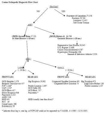
Orthopedic diseases in dogs account for ~22% of all small animal diagnoses, based on the Veterinary Medical Data Base. The canine orthopedic diagnostic flow chart presented here (Figure) is a useful tool for diagnosing orthopaedic diseases in dogs by guiding the clinician to the most likely and logical group of diagnoses.
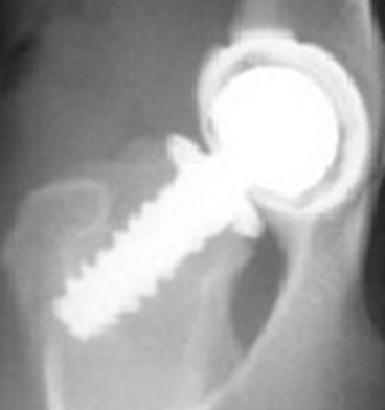
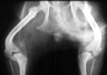
CHD has been called inherited, a developmental disease, and most accurately in the author's opinion, a "moderately heritable disease". CHD is a multifactorial disease with part of its cause being from genetic influences (estimated at 25%-80%) and part from environmental influences.
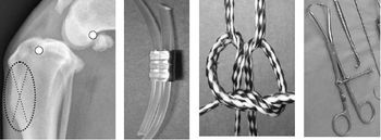
Conservative treatment of CCL rupture is the same as treatment for DJD: exercise to build muscle, weight control and medications such as NSAID's and Adequan. Surgery is the most effective treatment for CCL tear if done before DJD is established. Conservative treatment is indicated if surgery is not chosen by the owner because of cost or other reasons.
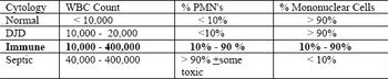
Synovial membrane lines all diarthrodial joints. Synoviocytes are macrophage like cells which phagocytize foreign materials and produce synovial fluid which contains the two lubricants hyaluronic acid and polysulfated glycosaminoglycans. The normal synovial membrane is a poor filter, allowing all components of blood into the joint fluid except cells, platelets, and large molecules such as fibrinogen.
OCD of the hock occurred bilaterally in 42% of the reported cases. The lateral trochlear ridge is involved in 25% of the cases and the medial trochlear ridge in 75% of the cases.
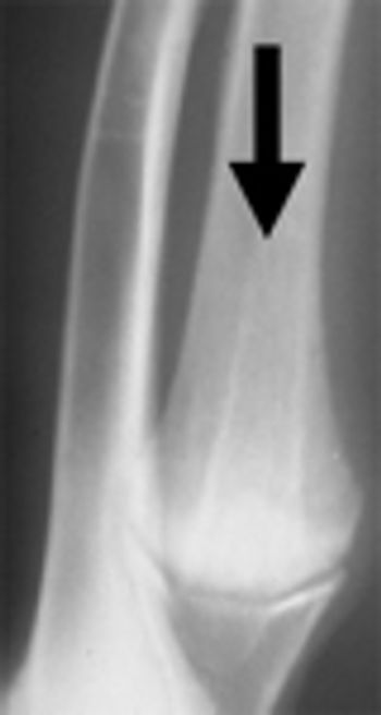
The best treatment for degenerative joint disease is prevention, by removing the inciting cause before DJD is established if at all possible (TPO, JPS, cruciate stabilization, patella stabilization, etc.) Irreversible changes of DJD (visible in the form or periarticular osteophytes) are present by 28 days after the cause is present.
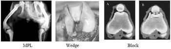
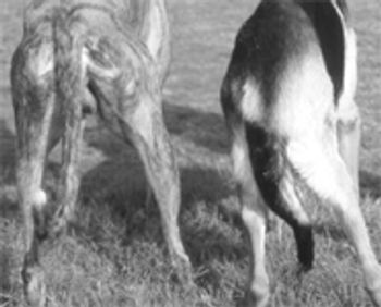
The patella is a type A (primary function is articulation) sesamoid bone located in the tendon of insertion of the quadraceps muscles. The origins of the quadriceps muscles are the proximal femur and immediately cranial to the acetabulurn (rectus femoris m.). The quadriceps m. follows a straight line, by necessity, to its insertion at the tibial crest.
Agenesis and malformation of the phalanges, metacarpal and carpal bones in utero is the basic pathology, and is occasionally accompanied by subluxation of the elbow.
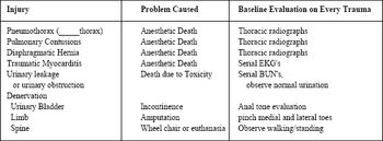
Thorax: Many dogs with pneumothorax (hemothorax, chylothorax, diaphragmatic hernia, etc) are able to ventilate marginally but adequately when at rest in a cage. The loss of lung/ventilation capacity may not become clinical for several minutes after induction of general anesthesia or during recovery.


