Dr. Clifford (Kip) Berry explains why radiographs are great for initially identifying the cause of vomiting in cats and dogs.
Dr. Clifford (Kip) Berry explains why radiographs are great for initially identifying the cause of vomiting in cats and dogs.
Dr. Clifford (Kip) Berry provides insight on how much training is required to master the ultrasound machine.
Abdominal ultrasound has taken on a "larger-than-life" position in diagnostic imaging in veterinary medicine for several reasons. First and foremost is that ultrasound is a non-invasive technique that can be used by the small animal practitioner for imaging the peritoneum, parenchyma of the abdominal organs and retroperitoneum.

Aside from ultrasound, a digital radiography suite is going to be the most expensive upgrade for the private practitioner in the realm of diagnostic imaging. However, this expense will be well worth the investment from a diagnostic imaging standpoint. After more than a century of film and film screen imaging, the backdrop of diagnostic radiology has changed from hanging films on view boxes to LCD monitors.
This brief overview of abdominal ultrasound is meant to be just that – an overview. If you are serious about ultrasound, you will take it upon yourself to review on the ultrasound references in physics such as that by Kremkau.
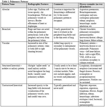
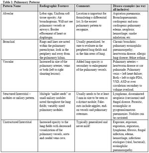
Pulmonary patterns have the bane of radiology since the beginning of time (1896 when x-rays were discovered). Pulmonary pattern recognition is the most difficult concept to teach and the most difficult and frustrating to learn, yet, the pattern itself is only part of the puzzle.

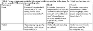
Mediastinal abnormalities, including cardiac disease, are common causes of clinical signs related to the thorax. By definition, the mediastinum is the midline potential space formed between the two pleural cavities and includes the medial portions of the right and left parietal pleura (also called the mediastinal pleural) and the space formed between these serosal membranes.
Thoracic radiography is still the most common first line assessment for diseases and conditions of the thorax. With the advent of digital radiography, a new interest in diagnostic radiology has emerged. However, even though certain artifacts are not an issue (e.g., processing, exposure), problems with inadequately positioned patients still exist.
Pulmonary patterns have the bane of radiology since the beginning (1896 that is). The most difficult concept to teach and the most difficult to learn, yet, the pattern itself is only part of the puzzle. The recognition that the disease is actual within the pulmonary parenchyma and not in the pleural space, extrathoracic structures or the mediastinum is the first step.
Aside from ultrasound, a digital radiography suite is going to be the most expensive upgrade for the private practitioner in the realm of diagnostic imaging. However, this expense will be well worth the investment from a diagnostic imaging standpoint. After more than a century of film and film screen imaging, the backdrop of diagnostic radiology has changed from hanging films on view boxes to LCD monitors.