
Anthony Pease, DVM, MS, DACVR, outlines how thoughtfully designed continuing education can help clinicians practice more efficiently without sacrificing quality.
Anthony Pease, DVM, MS, DACVR, outlines how thoughtfully designed continuing education can help clinicians practice more efficiently without sacrificing quality.

Worried that DR will become obsolete as soon as you make the purchase? Dont worryjust like fine diamonds, this technology holds value for years to come.
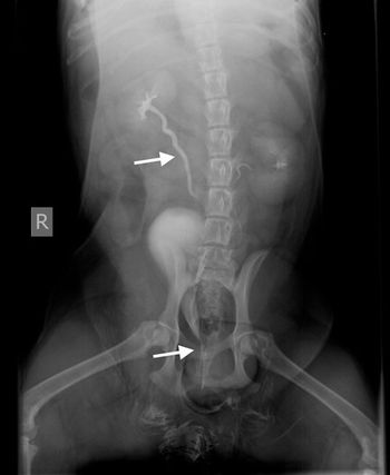
Veterinary radiologist Dr. Anthony Pease explains why contrast agents are a quick, easy way to examine the urinary tract
Veterinary radiologist Dr. Anthony Pease's thoughts on this quick and easy form of assessment.
Weird tales for the world from veterinary imaging.
Make more money, build your career and get props for being imaging wizard in your practice.
The use of computed tomography (CT), though not readily available at every institution, is becoming a more widespread modality for use in the small animal patient. Computed tomography affords a rapid evaluation of skeletal images with a small slice thickness that can be as small as 0.625.
Abdominal radiographs are a rapid, readily available method to give an overview of the abdomen. Though most people believe ultrasound is the new modality of choice for abdominal evaluation, the limitations of ultrasound not being able to penetrate gas as well as the technical ability and time to acquire images still make abdominal radiographs a great first modality in the patient with acute abdominal pain.
The explosion of magnetic resonance imaging (MRI) has revolutionized neuroimaging. However, other modalities still are valuable when looking at the central nervous system. These include ultrasound and computed tomography (CT). Radiographs have limited usefulness due to the superimposition of structures associated with the cranial vault and vertebral column.
The use of radiography to examine the abdomen is full of complications. Radiographs are very good at determining the difference between bone and gas, but soft tissue and fluid are the same opacity. When dealing with intra-abdominal lesions, the main goal is to differentiate one soft tissue mass from a normal soft tissue structure from abdominal fluid. Ultrasound uses high frequency sound waves to accomplish what radiographs cannot.
Ultrasound examinations are becoming routine standard of care in veterinary medicine. Due to the non-invasive nature of the modality, the general affordability of the hardware and the growing amount of continuing education to train general practitioners and veterinary technicians on the technical aspects of scanning, ultrasound is becoming the modality of choice, especially when examining the abdomen.
The evaluation of the musculoskeletal system is difficult due to the numerous soft tissues as well as the bone structures involved. Rapid assessment of the bone structure is routinely performed using radiographs; however, the subtlety of disease and joint compared to bone pathology can be confusing.
Diagnostic imaging has seen a huge technology shift in the last 10 years. Modalities that were not accessible to the small animal patient, such as magnetic resonance imaging, are now considered the modality of choice for neurologic examinations. This technology shift has caused a lot of confusion as well as questions about what modalities are used for which diseases and why.
November 1st 2010
November 1st 2010
November 1st 2010
November 1st 2010
November 1st 2010
November 1st 2010