










Urolithiasis is common in the cat, causing morbidity and, occasionally, mortality. Diagnosis is based on clinical signs, imaging, gross physical characteristics of the uroliths, and quantitative analysis of the urolith.
Urolithiasis is common in the cat, causing morbidity and, occasionally, mortality. Diagnosis is based on clinical signs, imaging, gross physical characteristics of the uroliths, and quantitative analysis of the urolith.
One of the common clinical scenarios that may present a diagnostic challenge is dogs or cats with urine retention without an obvious cause. The fundamental question in these cases is: "Does this patient have functional urinary retention or mechanical urinary obstruction?"
It's not enough to take radiographs if you don't take the views that you need to provide essential information. A series of radiographs will be reviewed that demonstrate the pitfall of omitting key information. Other examples of omission of key information will also be examined.
Diagnosis of urolithiasis may be aided by historical information such as the previous occurrence of uroliths or owner observation of symptoms compatible with the presence of uroliths. Predisposition to urolithiasis may be indicated by case signalment or pre-existing medical conditions favoring urolith formation.
The most common cause of feline lower urinary tract disease is idiopathic cystitis. Feline idiopathic cystitis, formerly called idiopathic feline lower urinary tract disease, is defined as a disease of undetermined etiology characterized by hematuria, dysuria, pollakiuria and possible urethral plug formation.

Protein can show up on a routine urinalysis for a variety of reasons.
In cats that are difficult to unblock, we use the soft portion of a 22-ga intravenous catheter and sterile saline solution in a 3-ml Luer lock syringe.
The recurrence of uroliths in cats is presumptively a common clinical situation; however, until now, no study has examined recurrence rates or the approximate time until recurrence for the main types of calculi found in cats.
Regarding the article "Incidental finding of renal agenesis in a cat," this condition may be a common finding in the Northeast.
Proteinuria is routinely detected by semi-quantitative, screening methods, like the conventional dipstick colorimetric test (very common) and the sulfosalicylic acid (SSA) turbidimetric test (less common). The dipstick test is inexpensive and easy to use. This test primarily measures albumin, however both the sensitivity and specificity for albumin are relatively low with the dipstick methodology.
Hyperthyroidism is one of the most commonly diagnosed diseases of the older cat. Geriatric cats with hyperthyroidism may also have concurrent chronic kidney disease (CKD). Systemic hypertension, proteinuria, and urinary tract infection (UTI) can be consequences of either hyperthyroidism or CKD.
Azotemia is defined as increased concentrations of urea and creatinine (and other nonproteinaceous nitrogenous substances) in the blood. The interpretation of serum urea nitrogen and creatinine concentrations as a measure of renal function requires a knowledge of the production and excretion of these substances.
Urinary calculi, or uroliths, are concretions of solid mineral and organic compounds that cause disease through direct trauma to the urinary tract and obstruction of urinary outflow. The urethral process is the most common site of obstruction in sheep and goats.
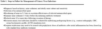
Most bacterial infections of the lower urinary tract respond quickly to antimicrobial treatment; however, urinary tract infections (UTI) associated with defects in the host immune system (complicated UTI) often fail to respond or recur after antibiotic withdrawal and can be a therapeutic challenge.
Chronic kidney disease (CKD) is a common problem that affects an estimated 0.5 to 7% of dogs. Radiographic signs of osteoarthritis (OA) occur in 20% of dogs. The majority of OA and CKD are acquired and both conditions are more prevalent in older dogs. Use of non-steroidal anti-inflammatory drugs (NSAIDs) has dramatically improved the quality of life for many dogs with OA.
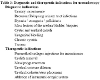
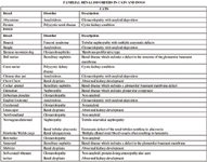
The clinical signs of many lower urinary tract diseases (LUTD) in dogs and cats are similar. Correct identification of the underlying disease process is critical to the development and implementation of a successful treatment plan.
Systemic hypertension in animals has largely been thought to be secondary to another disease (e.g., renal disease and endocrinopathies), as opposed to idiopathic (primary or essential). This has recently been called into question. For example, in a report of 69 hypertensive cats, seen at North Carolina State University (NCSU) for ocular disease, revealed that at least 17%, and possibly as many as 50%, of cats had no identifiable cause for their systemic hypertension.

Chronic kidney disease (CKD) is a common problem that affects an estimated 0.5 to 7% of dogs and 1.6 to 20% of cats. Nephron damage associated with CKD is usually irreversible and can be progressive. Renal failure results when three-quarters or more of the nephrons of both kidneys are not functioning.
Successful management of urolithiasis in dogs and cats depends upon the removal of existing uroliths and preventing their recurrence. Traditionally, uroliths have been removed via surgery.
Acute kidney injury (AKI) often results from ischemic or toxic insults and usually affects the most metabolically active tubular portions of the nephron. If the ischemic or toxic insult is severe enough, acute renal failure (ARF) may result. In many cases, AKI and ARF inadvertently develop in the hospital setting in conjunction with diagnostic or therapeutic procedures.
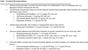
Micturition is controlled by a combination of autonomic and somatic innervation. Sympathetic innervation to the bladder via the hypogastric nerve is composed of preganglionic fibers exiting the lumbar spinal cord from the L1-4 spinal cord segments and synapsing in the caudal mesenteric ganglion.
The normal feline lower urinary tract has a number of defence mechanisms against infection. These include normal micturition (e.g., frequent and complete voiding), normal anatomy (e.g., length of urethra), uroepithelial mucosal barriers, the antimicrobial properties of normal urine (e.g., high specific gravity and osmolality) and a normal immune system.
Lower urinary tract disease in cats was described as early as 1925. Over the years, the terms "feline urologic syndrome" (FUS) and "feline lower urinary tract disease" (FLUTD) have been used to describe the group of clinical signs related to problems voiding. However, these descriptive terms do not identify the underlying etiology.
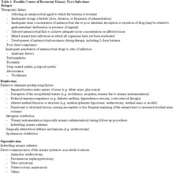
Urinary tract infection is the most common infectious disease of dogs, affecting as many as 14% of dogs over the course of their lifetime. The majority of these urinary tract infections (UTIs) are benign and respond readily to antimicrobial therapy.
Struvite and calcium oxalate (CaOx) uroliths are the most commonly reported uroliths in cats. In the last 25 years, dramatic change in the prevalence of different urolith types has occurred. Until the mid-1980s, struvite uroliths made up 78% of submissions to the Minnesota Urolith Center (MUC).