




















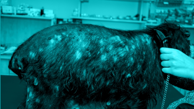

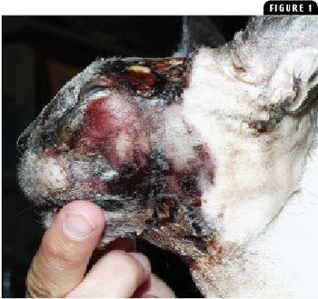
Feline AMP is an uncommon infectious disease of dermal tissues and subcutaneous fat.
Feline AMP is an uncommon infectious disease of dermal tissues and subcutaneous fat.
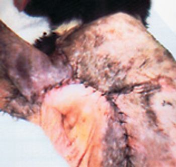
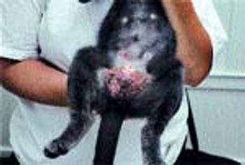
A 6-year-old, 11.2-lb (5.1-kg), neutered male domestic shorthaired cats was presented to South Athens Animal Clinic for evaluation of a large cutaneous wound.

In my multi-specialty, referral-based veterinary hospital in the heart of San Francisco, with all our many bells and whistles, including access to numerous specialists within our clinic (internal medicine, surgeon, radiologist, dentistry, holistic, oncologist, acupuncturist and ophthalmologist), I still continue to feel very frustrated with a common skin disease: canine pyoderma.
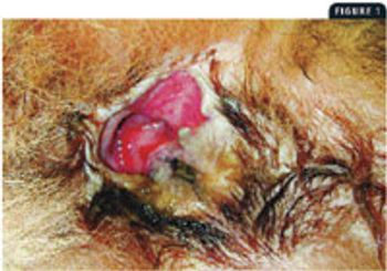
How much success have you had using topical tacrolimus as a single agent once or twice a day to treat perianal fistulas?

Cyclosporine is an immunosuppressive drug that suppresses T-helper and T-suppressor cells and inhibits interleukin-2.
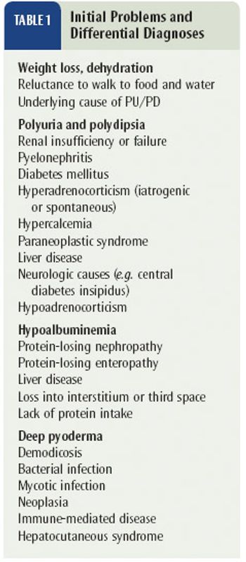
A 14-year-old, 41-lb (18.5-kg), spayed female collie-mix presented for evaluation of intense pruritus and gradually worsening skin disease of more than one year's duration.
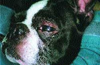
Previously, differentials of feline facial pruritus were discussed to include ectoparasites such as flea allergy, otodectes, Notoedres and cheyletiella, food allergy, atopy including possible food storage mite allergy and demodicosis. Less common differentials include infections such as dermatophytosis, viral, and bacterial pyoderma, Malassezia dermatitis, Pemphigus foliaceus (PF) and idiopathic facial dermatitis of the Persian cat. Clinically, many of these diseases appear similar including Pemphigus foliaceus and bacterial pyoderma which can be difficult to differentiate both clinically and histopathologically.
We have pretty much resolved ourselves to pigeonholing our pruritic dogs into the categories of atopy, food allergy and ectoparasites as a reason for their itching.
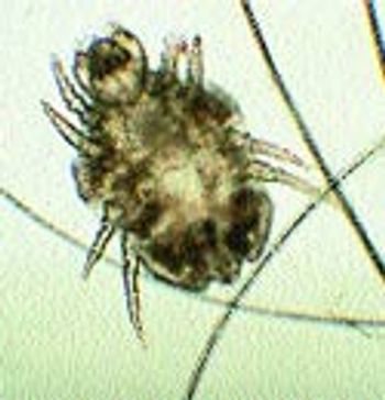
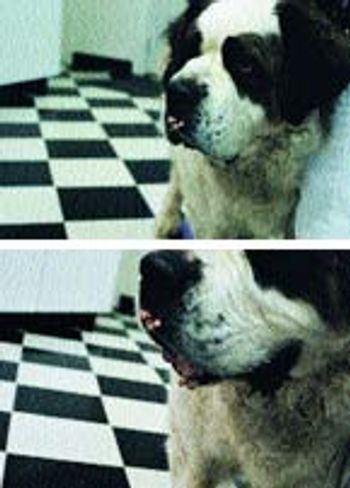
For those nonseasonal pruritic patients where food elimination trials haven't been helpful and steroids seem to help or those patients with nonseasonal recurrent otitis or pyoderma, an allergy to house dust mites or food storage mites should be considered. Dust mites are the leading cause of allergy/asthma in humans. In dogs, it appears to exceed flea allergy as the most common intradermal skin test reaction. An estimated 30-80 percent of atopic dogs and cats skin test positive to dust mites (Photo 1). Of the two types of dust mites, Dermatophagoides farinae and Dermatophagoides pteronyssinus, cats tend to be allergic to
Test your dermatology skills on the following cases. What would you do first? Which diagnostics would yield the most information yet not break your client's wallet? Are you seeing an unusual disease or more commonly, an unusual manifestation of a common disease?
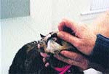
Q. I have a cat with a rodent ulcer that used to respond to steroid injections. Is there anything new for this?
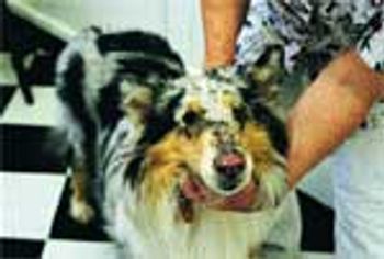
In my last article, I discussed skin diseases that result from not enough sun exposure, i.e. seasonal flank alopecia or light responsive alopecia. With the coming of summer, it is timely to offer attention to skin diseases that are exacerbated by sunlight.
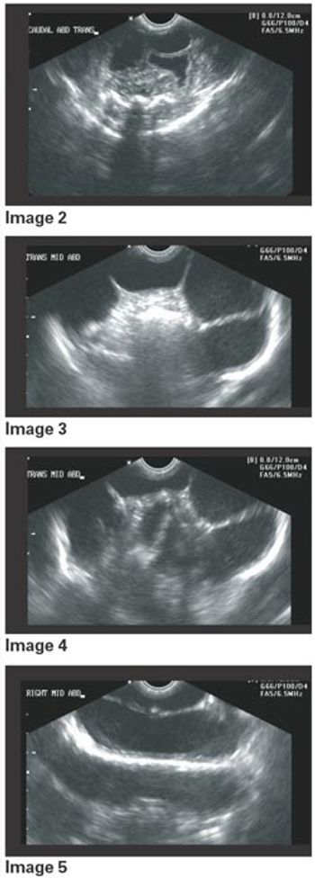
Editor's Note: In our ongoing telemedicine series, Dr. Johnny Hoskins presents medical case studies. The format is heavily focused on radiology and ultrasonography and details complicated, yet fairly common cases most veterinarians will be exposed to in practice.
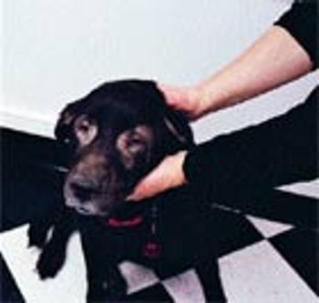
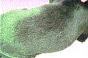
Coming off of a long, dark winter as we are in the Midwest, we're confronted with a unique disease seen in certain breeds of dogs. Light responsive alopecia or seasonal flank alopecia is most often seen at this time of the year.
At one time or another, we have all made the same mistakes when working up a dermatology case. To help us all save time and get the most information with the least amount of work, I thought I would address what in my referral practice appear to be the most commonly made mistakes when working up a dermatology patient.
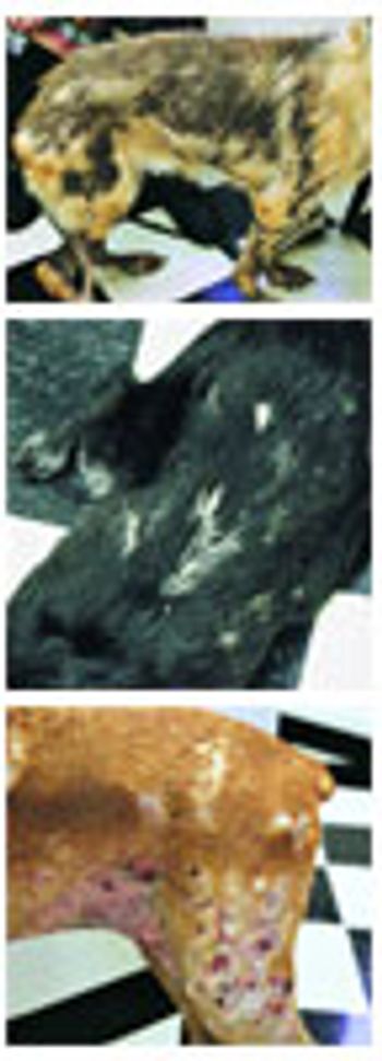
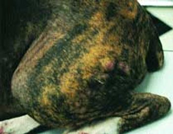
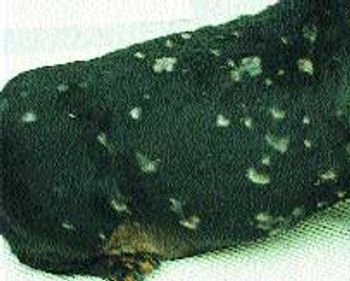
"Why does my dog have dry skin? Didn't I wash off all the shampoo? Am I bathing him too much? Does he need a conditioner?" We have all been asked these questions many times. With the winter months upon us for those in the colder areas of the country, the low humidity often causes humans to have "dry skin". This may be true for our canine patients as well, however dry skin in dogs may be the result of several underlying diseases (Photo 1).
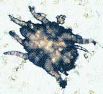
Canine scabies is often missed (reportedly 70 percent of the time) and should always be considered.
Dr. Alice Jeromin identifies skin conditions that can serve as early warning signs of internal disease. Using these clues practitioners can help prolong the lives of their patients.
All of us, at one time or another, were probably guilty of treating one of our sarcoptic mange patients as an allergic patient. It is the perfect example of a patient with the same clinical appearance and symptoms of two diseases: atopy vs. sarcoptic mange.
In DVM Best Practices on Feline Medicine (May, 2002), I wrote about feline ear mites and dermatophytes, two common infectious diseases often seen in feline practice.
Dr. Michele Rosenbaum outlines diagnostic and clinical management options for two of the most common infectious diseases seen in feline patients.
As mentioned in the first article (Feb. 2002) of this series, the presentation of the pruritic dog can be frustrating for the veterinarian because of the number of possible differential diagnoses.
When clients bring in their pets to have growths removed or wounds examined, we have the clients mark the problem spots on an anatomy chart. The chart makes it easy for us to locate all the lumps and lesions and is a great alternative to drawing on the animals with a marker. --Sage Olson, receptionist Kensington, Conn.
One of the most frustrating and time-consuming problems in everyday veterinary medicine is the presentation of the dog with pruritus.
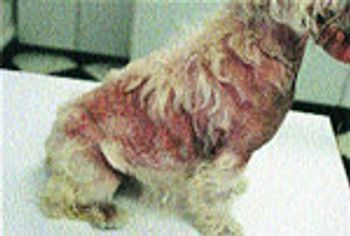
I often get referrals of patients on immunotherapy that "aren't doing well" or hear from the owner that "those shots never helped so we stopped them."