














The role thoracic radiography should play in determining the severity of lung disease in dogs.
The role thoracic radiography should play in determining the severity of lung disease in dogs.

Traumatic thoracic injuries are prevalent in small animals, particularly in dogs. The most common causes of thoracic trauma are motor vehicular accidents and bite wounds. Other possible, although less common mechanisms include gunshot, knife wounds or being kicked by a larger animal (horse/cow). Injuries may range from mild to life threatening.
Tracheal stenting has been recently re-introduced as an approach to management of severe tracheal collapse. Severe tracheal collapse is life-threatening and while extra-thoracic collapse may be amenable to extra-luminal prosthetic rings, intra-thoracic collapse is not.
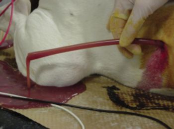
The purpose of this lecture is to review the management of pyothorax in cats and dogs. Pulmonary infection can result from bacterial, viral, fungal or protozoal infection, however, pyothorax is almost uniformly bacterial. The pleural space has a small amount of fluid normally (~ 5 ml) which serves as lubrication for the pulmonary parenchyma.
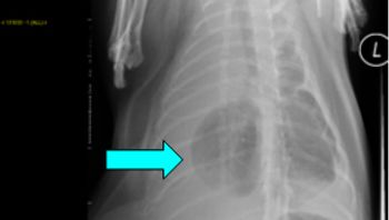
Pneumothorax is defined as free air in the pleural space. Normal intra-pleural pressure is about - 5 cmH20, which means that in order to equilibrate pressures, air from either the atmosphere, or the lung will equilibrate rapidly with the pleural space. Pneumothorax can be further characterized as traumatic, spontaneous and iatrogenic.
Respiratory distress is common and challenging. Cats often compensate well for pulmonary diseases, and some conditions can rapidly fulminate. Dogs are often more "honest" although they can decompensate rapidly as well. It is crucial to balance the equal goals of limiting stress on the patient with respiratory distress, and to work to identify the specific cause of the distress so that appropriate therapy can be provided.
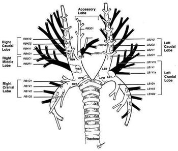
Definitive diagnosis of pulmonary disease remains elusive at times. Cytological or histopathological samples are useful to help better classify the underlying cause as well as determine both prognosis and treatment course. Thus, it is prudent for the criticalist to have a strong grasp of the various techniques and options available for sampling.
Upper airway diseases/obstruction are relatively common causes of respiratory distress in dogs and cats. However, because lung parenchymal diseases are more frequently observed, upper airway problems may be overlooked. In order to fully appreciate upper airway disease, it is essential to be familiar with the structure, function, and common abnormalities.
Feline bronchial disease goes under a multitude of names reflecting the considerable heterogeneity in anatomic locations as well as etiologies that may be involved.
Herpesvirus 1 (rhinotracheitis; FHV-1) and calicivirus (FCV) are the most common viral causes of sneezing and nasal discharge in the cat. If oral ulcers are present, calicivirus is most likely. If corneal ulcers are present, herpesvirus 1 is most likely. FHV-1 has now also been associated with chronic stomatitis, facial dermatitis, and endogenous uveitis. Viral rhinitis with or without secondary bacterial infection can be recurrent.
Dogs with this condition are often characterized as having a "smoker's cough."
In cats, bronchitis and asthma can be difficult to distinguish.
Why acute lung injury and ARDS may sneak up on you.
Advice on one rule-out: lung parasites.
This form of therapeutic intervention may be just the answer for this disease that affects up to 5% of the pet cat population.
Is it herpesvirus? Is it a bacterial infection? Drs. Little and VanVranken discuss how to handle these cases.
The history of canine influenza virus began with the identification of infections in racing greyhounds, directing research that defined canine influenza virus (CIV), a variant of equine influenza virus with a unique genetic signature capable of being transmitted from dog to dog.
Buckinghamshire, UK - A new research project shows that genes classified as mucins may regulate mucous proteins and change over the course of equine respiratory disease.
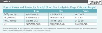
An introduction to the principles of clinical blood gas analysis and interpretation--including two case examples.
Feline bronchopulmonary disease (FBPD), often referred to as "feline asthma" actually encompasses a group of common, but poorly understood, airway diseases. It is estimated that bronchopulmonary disease affects 1% of the general cat population and > 5% of the Siamese breed. Cats of any age can be affected and there is no clear gender predisposition.
A large number of disorders (infectious, non-infectious inflammatory, immune-mediated, neoplastic etc.) can affect the bronchopulmonary tree in dogs. A comprehensive review of each disorder is beyond the scope of this presentation. Rather we will review key clinical points about the diagnosis and management of canine chronic bronchitis and pulmonary fibrosis.
Cats who cannot breathe are the most fragile patients we treat each day. Cats tend to be more compromised on presentation as they hide their breathing issues better from their owners. It is important to balance diagnostic procedures with therapeutic intervention so that these cats can be quickly stabilized and effectively treated.
A variety of disorders can affect the upper respiratory tract of cats; while the feline upper respiratory infection complex is one of the most common URT disorders, this lecture will focus more on more complex disorders such as chronic rhinitis, nasopharyngeal polyps and nasopharyngeal stenosis, as well as feline laryngeal disease.
Traumatic thoracic injuries are prevalent in small animals, particularly in dogs.
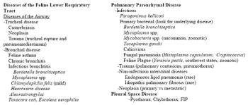
If the cat is presented for labored breathing questions should be directed to assist in differentiating upper from lower respiratory disease as some owners perceive stertorous breathing in a cat with upper respiratory disease as a form of labored breathing. This could be misleading to a clinician.