
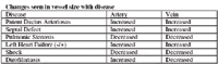
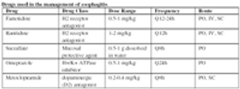
Esophageal foreign bodies and esophagitis have the potential, if not identified and treated, to cause esophageal strictures or megaesophagus, which can be more difficult to treat.
Esophageal foreign bodies and esophagitis have the potential, if not identified and treated, to cause esophageal strictures or megaesophagus, which can be more difficult to treat.

A review of the indications for radiography and ultrasonography.
Primary gastrointestinal (GI) neoplasia is an important differential diagnosis for a dog or cat with vomiting or diarrhea, especially chronic vomiting or diarrhea, anorexia and weight loss, particularly animals that are middle-aged and older.
Diagnostic ultrasonography has, more recently, been applied to the assessment of other less traditional musculoskeletal problems such as evaluation of bone, joints, muscle and nerves.
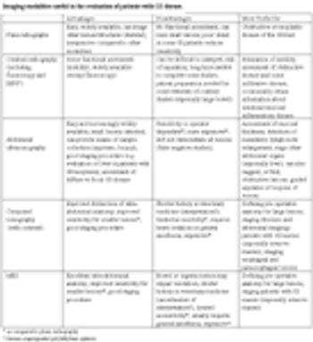
The small animal clinician has a number of imaging options available for the evaluation of dogs and cats with gastrointestinal tract (GI) disease.
Do not buy used "very old" equipment, but equipment that is one generation old is often the best value.
Inflammatory bowel disease is a somewhat loosely defined term that describes chronic gastrointestinal (GI) tract signs in dogs and cats, especially weight loss, vomiting and diarrhea.
Diagnostic imaging is an underutilized resource in herpetological medicine.
Ultrasonography is invaluable in the diagnosis of the cause of colic in horses.
Thoracic ultrasonography yields information about the lung and pleural cavity heretofore unavailable to the veterinarian.
Many of the exotic species we deal with have evolved to mask their illness to avoid predation.
The protein-losing enteropathies (PLE) comprise a collection of intestinal, usually small intestinal, diseases typically associated with weight loss, hypoproteinemia caused by hypoalbuminemia or panhypoproteinemia, and variable signs of weight loss, vomiting and diarrhea.
Take as many views as you need to in order to define a lesion or feel confident the lesion can not be defined using this technology.
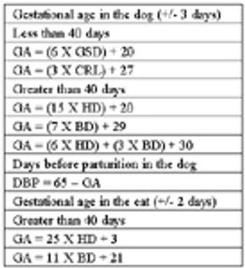
In the bitch and queen, documented abnormalities of the estrous cycle, pregnancy and the periparturient period, and even disorders of the residual reproductive tract in ovariohysterectomized females, call for ultrasonographic evaluation of the uterus and ovaries.
The normal uterus is best located by scanning transversely between the urinary bladder and the colon.
The use of ultrasound as a tool in canine and feline reproduction has expanded from its initial role in early pregnancy diagnosis to its current use in the approach to clinical reproduction (obstetrics, infertility, urogenital disorders and pediatrics).
Abdominal ultrasound provides valuable clinical information about the peritoneal cavity, great vessels, abdominal viscera and lymph nodes obtained in a noninvasive fashion, with no confirmed adverse biologic effects, and usually not necessitating sedation or anesthesia.
Pediatric patients are commonly presented to the veterinarian because of signs referable to the abdominal cavity due to congenital anomalies, dietary indiscretion, parasitic infestation and infectious disease.
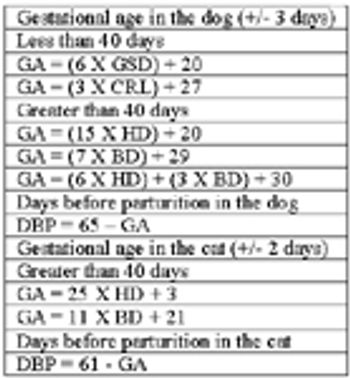
Diagnostic ultrasonography has become an important component of small animal theriogenology since its introduction to practice in 1978.
Generally, any evidence of change in reproductive performance or of genitourinary disorders detected on physical examination or laboratory analysis indicates the need for ultrasound evaluation in the male dog and cat.
One of the first steps in switching to digital radiography is deciding what type of system to purchase.
National Report - Pet owners are shopping for price, veterinarians say, and it's impacting general practice and referrals.
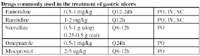
Are contrast studies still needed as diagnostics in vomiting patients?
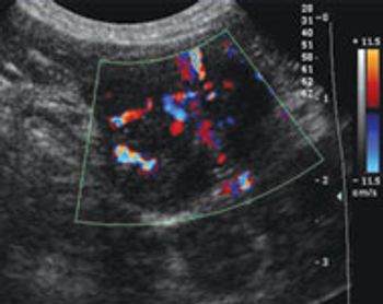
One of the strengths of ultrasound imaging is its ability to resolve very small soft-tissue structures.
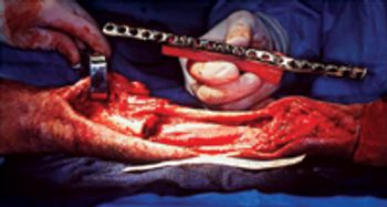
Amputation remains the standard of care to address most primary tumors of the appendicular skeleton. But in certain cases, a limb-sparing surgical procedure may be an option.
