


Anatomic and physiologic considerations are the basis for proper diagnostic techniques. We will discuss basic diagnostic procedures and relative pharmacological consideration to enhance the ophthalmic examination.
Ophthalmology
Latest News
Advertisement
CME Content
Advertisement
Anatomic and physiologic considerations are the basis for proper diagnostic techniques. We will discuss basic diagnostic procedures and relative pharmacological consideration to enhance the ophthalmic examination.
Dogs and cats are very commonly presented for management of acute or chronic wounds in veterinary practice. The goal of wound management is to accelerate the healing process and not to interfere with it.
The "red eye" may present for many different reasons. Periocular trauma, conjunctival hyperemia, corneal vascularization, intraocular hemorrhage, and detached subalbinotic retinas may all lead to the complaint of a "red eye". Examination of the eye should lead to localization of the abnormality and standard ophthalmic testing should be performed, including schirmer tear tests, fluorescein staining, and intraocular pressures.
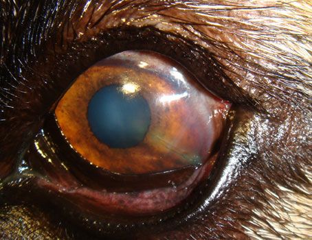
Early recognition of glaucoma is essential in managing this disease and preventing the natural outcome, which is a painful and blind eye. Recognition is preceded by having a suspicion for glaucoma, and various signs, including fixed and dilated pupils, engorged episcleral vessels, and a hazy cornea, should heighten this suspicion.
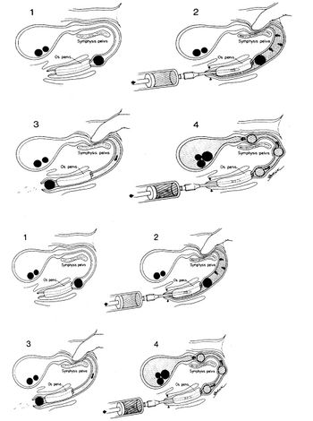
Cryptorchism is defined as the failure of one or both testicles to descend into the scrotum. The cryptorchid testicle can be located anywhere along the path from the area of fetal development of the gonads (just caudal to the caudal pole of the kidney) to the subcutaneous tissue between the external inguinal ring and the scrotum.
Eyelid function is important in maintaining the health of the cornea and globe. Eyelids distribute tears over the corneal surface, remove foreign bodies from the surface of the eye, control the amount of light entering the eye and protect the globe from trauma.
Pertinent ophthalmic anatomy for veterinarians in private practice is reviewed from the outside in, as are related diagnostic tests and pertinent diseases. In order, the orbit, eyelids, third eyelid, tear film, nasolacrimal drainage system, cornea and sclera, lens, uveal tract (iris, ciliary body, choroid), iridocorneal angle and aqueous dynamics, vitreous, retina, optic nerve, and visual cortex are reviewed.
Portosystemic shunt (PSS) is an abnormal vessel that shunts portal blood from the splanchnic circulation to flow directly to the systemic circulation by passing the liver. Toxins, hormones, nutrients, escaping bacteria, and exogenous drugs also bypass the liver resulting in hepatic encephalopathy (HE).
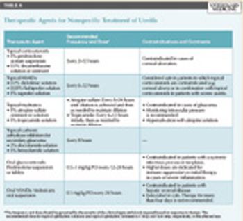
Uroliths can be found in the renal pelvis, the ureter, the bladder, or the urethra. The most common locations are the bladder and the urethra. A higher incidence of calcium oxalate has been reported in the ureter and kidney in cats.
A thorough fundic evaluation is important in diagnosing retinal disease. At the beginning of the exam the menace response, dazzle reflex and the pupillary light reflexes should be assessed. Abnormalities on the neuro-ophthalmic portion of the exam may augment interpretation of fundic findings.
True ophthalmic emergencies commonly seen in small animal practice include acute primary glaucoma, anterior lens luxation, traumatic globe proptosis, and progressive deep corneal ulceration. It is important that the general practitioner be able to recognize these sorts of emergencies.
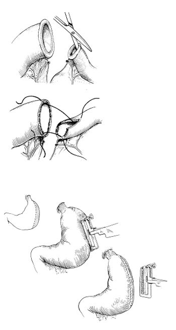
Gastrointestinal surgery is performed very commonly in small animal for biopsy, removal of a foreign body, upper gastrointestinal bleeding, resection of a necrotic segment of intestine, resection of necrotic portion of the stomach, and resection of a neoplasia.
The lens is composed of crystalline fibers specifically arranged to allow light rays to transilluminate through the lens to the retina. The lens focuses light rays on the retina.
The laryngeal functions are to regulate airflow, voice production, and prevent inhalation of food. If the intrinsic muscles and/or the nerve supply of the larynx are not normal laryngeal functions are compromised.
Corneal surgical techniques available for use in veterinary medicine are reviewed. Surgeries discussed include linear grid keratotomy, multiple superficial punctate keratotomy, diamond burr superficial keratectomy, thermokeratoplasty, keratectomy, conjunctival graft placement (pedicle, island, bridge, advancement, etc.), corneoconjunctival transposition flaps, penetrating and lamellar keratoplasties (corneal transplants), biosynthetic graft placement (A-cell, BioSist), and amnion graft placement.
A 9-year-old 46.2-lb (21-kg) spayed female mixed-breed dog was presented to the ophthalmology service at Colorado State University for evaluation of incisional infection and delayed healing after bilateral entropion surgery.
National report -- The third annual National Service Dog Eye Exam Event is slated for May but registration begins April 1.
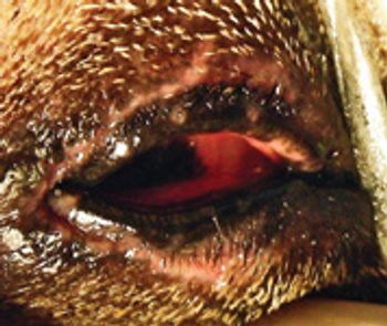
Nonspecific therapy for feline anterior uveitis includes topical mydriatics, corticosteroids, and nonsteroidal anti-inflammatory drugs (NSAIDs).
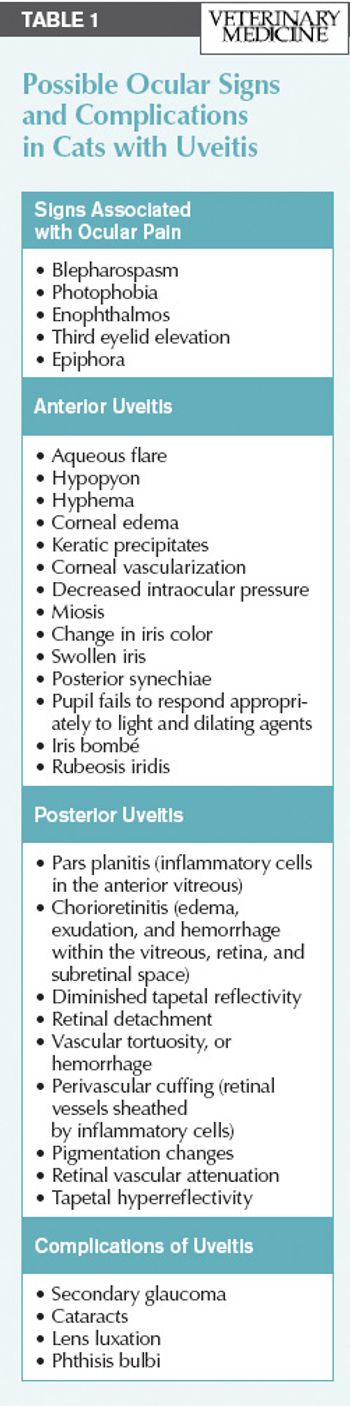
Uveitis is a common and painful ocular disease in cats that can eventually lead to blindness.
Patients that present for ophthalmic surgery are often geriatric or pediatric, painful and anxious, and may present as emergency cases.
At what age do cats open their eyes?
In this text, I will touch on a few select ocular emergencies that are deemed dire if vision or the globe is to be saved.
The normal cornea is clear, and any alteration in clarity signifies pathology.
Anatomic and physiologic considerations are the basis for proper diagnostic techniques.
Advertisement
Advertisement
Trending on dvm360
1
Q&A with an emergency veterinarian on critical care and common ER misconceptions
2
Creatine for veterinary professionals (Part 1): What it is, how it works, and why you should care
3
AAVSB tapped to develop VPA licensing examination
4
FDA launches pilot to boost US manufacturing of animal drugs
5