


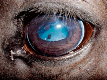
What condition are these ocular changes consistent with?
What condition are these ocular changes consistent with?
Most sources state that the reference range of Schirmer tear test values is 15 to 20 mm/min in most species.
Treating the primary cause is indicated in most conjunctivitis cases.
A look at how several antifungal agents and delivery vehicles affect equine keratocytes.
When suturing after a wedge resection from an eyelid margin, it can be challenging to keep the suture ends from irritating the eye.
The results of a University of Wisconsin randomized controlled trial assessing the efficacy of retrobulbar analgesia on the control of postoperative pain in dogs undergoing enucleation was recently published.

What are the signs of melting corneal ulcers?
How to make sure surgery to correct this problem doesn't lead to ectropion.
This diagnostic test is often difficult and stressful for the pet, owner, and veterinarian.
Corneal disease and damage resulting in cloudy, blue eyes has many causes.
When presented with exotic species with ocular disease it is important to remember the concept of "the same and different". Eye are eyes regardless of the species and many of the conditions we are presented with in pocket pets and other exotic species are in many ways the same as those we have come to recognize in the more usual domestic species and can be diagnosed and treated empirically in the same way.
Pediatrics, for the purpose of this lecture, will be considered conditions that are genetic, congenital, and early developmental and acquired. They are common in the first year of life. To have the best understanding of many of the genetic and congenital diseases, a review of embryology will be necessary, and we will further highlight embryology as we discuss many of these conditions.
Many ocular conditions seen in cats are identical to those in other domestic species however there are eye diseases which are only seen with any frequency in cats (eyelid agenesis, diffuse iris melanoma) or which have common and unique presentations in the cat compared with other species (immune mediated uveitis and ocular eosinophilic disease).
A good ocular examination begins with a complete medical history. The saying goes that the eyes are the window to the soul – to the ophthalmologist they are often a window to illness elsewhere in the body. The general medical history should be scrutinized starting with signalment and work/play/housing environments.
The eyelids often have increased flaccidity and laxity in advanced age. This may result in entropion, often of the lateral aspect of the upper eyelid. Loss of orbital fat pad may result in enophthalmos and protrusion of the third eyelid. This too can lead to entropion, typically of the lower lids.
The SCCED represents a specific unique type of corneal ulcer that is frustrating for veterinarians and clients alike. They are chronic, superficial, non-infected, and present with the patient minimally to severely painful. Most are characterized by a superficial erosion of the corneal epithelium with loose epithelial edges and variable corneal vascularization.
Distichiae are aberrant hairs that arise from the meibomian glands and exit through the meibomian gland opening in the eyelid margin. Distichiasis occurs commonly in many breeds of dogs. In some breeds the hairs cause virtually no clinical signs (e.g., Cocker Spaniels), whereas in other breeds they can result in epiphora, corneal fibrosis and vascularization, and occasionally corneal ulceration.
A good ocular examination begins with a thorough medical history. The saying goes that the eyes are the window to the soul – to the ophthalmologist they are often a window to illness somewhere else in the body. Start with the basics; signalment, use, as well as housing, work, and turnout environments.
When we examine the posterior segment of the eye we tend to think that we are looking for problems in the retina. I reality we are seeing the vitreous humor, the neural retina and optic nerve, retina pigment epithelium, the vascular coat lining the back of the eye – the choroid, and the outer fibrous coat of the eye – the sclera. The fundus is the area including all of these structures as seen by ophthalmoscopy through the pupil.
Uveitis in the horse can be divided broadly into two forms – equine recurrent uveitis and non-recurrent/progressive uveitis
Corneal disease is the most common reason for ophthalmic examination in the horse. Many corneal lesions are initially due to trauma.
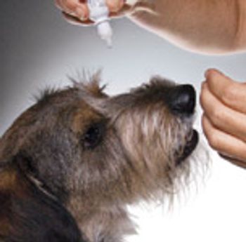
When administering eye drops to a dog, hold a treat above the dog's head, causing it to look up.

This online on-demand course reviews various color changes in the cornea that the clinician may observe in patients with ocular disease. (1 CE credit)
This online on-demand course reviews the diagnosis and management of ocular emergencies. (1 CE credit)
This online on-demand course reviews the diagnosis and management of ocular emergencies. (1 CE credit)