


Pulmonary hypertension (PH) is defined by a systolic pulmonary artery pressure greater than 25 mmHg. The incidence of PH is difficult to define due to lack of clinical awareness, non-specific clinical signs, and difficulty in confirming the diagnosis.
Cardiology
Latest News
Advertisement
CME Content
Advertisement
Respiratory abnormalities are relatively common in cats, who may suffer from a wide range of disease processes. An initial triage step in the management of a dyspnic cat is to determine whether the dyspnea is cardiogenic or due to extracardiac (primary pulmonary) disease. This task is in no way a simple one, as cats often have non-specific history and physical examination abnormalities.
Tricks to handle the adverse effects: never with- hold water, always have water available or there will be much more significant dehydration and azotemia.
Echocardiography has emerged as the most valuable non-invasive tool for evaluation of cardiac structure, function, blood flow patterns, and has greatly diminished the need for diagnostic cardiac catheterizations and angiocardiography in many cases. Echocardiography is one tool for evaluation of the cardiac patient, but should be used in conjunction with other diagnostic tests including thoracic radiography and electrocardiography for a global assessment of the patient.
Although there are highly sophisticated and advanced diagnostic modalities in cardiology, the basic technique of a good cardiovascular examination is still an essential fundamental element of the cardiovascular workup. Other basic diagnostic modalities that are readily available in most practices include thoracic radiographs, electrocardiography, and blood pressure measurement.

This online on-demand archived Webinar will highlight the importance of differentiating between heart disease and heart failure and will discuss the impact on diagnosis, therapy and monitoring your feline patients. (1 CE credit)
This online on-demand archived Webinar will introduce you to a classification scheme for heart disease and heart failure and will discuss the clinical significance of signalment, history and common cardiac diagnostic tests to appropriately stage dogs and cats with heart disease. (1 CE credit)

What is the aim of this heart disease treatment?
One of the biggest risk factors for this condition in cats.
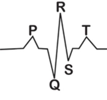
Technicians aren't all destined to be cardiologists. But all technicians should understand what a normal cardiac rhythm looks like and how it's generated.
New guidelines give veterinarians a tool for managing these cases in dogs.

Does the obesity paradox in people occur in cats as well?
Blood pressure measurement is often not a routine part of small animal practice. This partially has to do with the equipment available to measure blood pressure as well as our patients (they tend not to hold still like we have to).
Cardiovascular (CV) diseases in cats include congenital malformations, acquired heart, and vascular disorders. Myocardial disorders or cardiomyopathies, the major cause of heart failure, thromboembolism, and persistent arrhythmias in cats, constitute the focus of this presentation.
Arrhythmias can be classified based on ECG analysis based on the heart rate (normal, bradyarrhythmias, tachyarrhythmias); anatomic origin of the rhythm disturbance (SA, atrial, atrioventricular, or ventricular); or electrophysiologic mechanism when evident.
Acquired heart diseases of dogs include chronic degenerative valvular diseases (endocardiosis), pericardial diseases, cardiac neoplasia, dilated cardiomyopathy (DCM), arrhythmogenic right ventricular cardiomyopathy (ARVC), pulmonary hypertension (PH), infective endocarditis, and heart rhythm disturbances, some of which represent primary electrical disorders and others that develop secondary to cardiac remodeling.
The technician should appreciate the definition and causes of CHF, as well as methods of evaluation. The drugs used to treat CHF should be understood, as well as related side-effects. This awareness improves both patient care and client communications.
The signs of pulmonary parenchymal vascular disease are not always obvious.
February is National Heart Month and a good time to save cats from cardiac disease.
What is the most common cause of pericardial effusion in dogs?
Is there an indicator for severity of cardiac disease in dogs?
Orlando -- Pioneering veterinary cardiologist Stephen Ettinger, DVM, Dipl. ACVIM received the 2011 Mark L. Morris Sr., Lifetime Achievement Award yesterday.
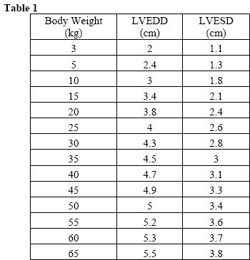
How can hydration status influence echocardiographic measurements?
An important aspect of diagnosis of this disorder in American cocker spaniels.
Anesthetizing a patient with cardiac disease requires a plan for the use of supportive measures to maintain adequate tissue perfusion. As in the case of left sided cardiac dysfunction patients, volume administration frequently is not an option to support blood pressure. In these cases, should a positive inotropic or pressor agent be indicated, the volume of the adjunctive agent required should be deducted from the volume of crystalloid administered to maintain a balanced hourly rate.
Advertisement
Advertisement
Trending on dvm360
1
Q&A with an emergency veterinarian on critical care and common ER misconceptions
2
Tornado (the dog) touches down at the emergency department
3
When veterinary partnerships work (and when they don’t)
4
Every 30 extra minutes under anesthesia raises complication risk in brachycephalic patients, underscoring the importance of peri-anesthetic management
5