

|Articles|February 1, 2006
Unlock dermatology secrets to realize treatment success
Author(s)Carlo Vitale, DVM, DACVD
When appropriately administered and not continued long-term, corticosteroids are very helpful and safe.
Advertisement
After finishing my residency at the University of California-Davis in 1994, I decided to remain on as a clinical instructor for an additional two years prior to starting my dermatology career in private practice. I really didn't know what I was getting into.
What I learned (and continue to learn) are wonderful and useful tidbits of knowledge that have allowed me to practice the highest quality of medicine. Here they are in no particular order.
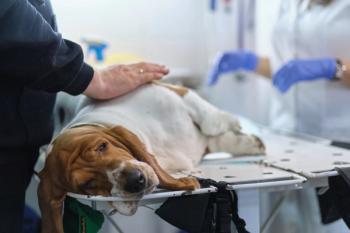
Treatment of recurrent pyoderma
Generally speaking, most cases of canine superficial pyoderma are cleared with one course of antibiotics, provided that it is an appropriate choice for Staphylococcus and the dosage and duration are adequate. The choices and dosages for antibiotics are listed below:
- Cephalexin 22mg/kg, orally BID
- Trimethoprim/sulfa 14mg/kg BID
- Ormetoprim/sulfa 55mg/kg once daily for 1 day, then 27.5 mg/kg once daily
- Cefpodoxime 5-10mg/kg once daily
- Enrofloxacin or ciprofloxacin 5 mg/kg once daily (I prefer 10 mg/kg, or higher)
- Marbofloxacin 1.25 mg-2.5 mg/kg once daily
- Clindamycin 15-20 mg/kg twice daily
- Lincomycin 22mg/kg twice daily
- Amoxicillin/clavulanic acid 20 mg/kg twice daily.
It's always important not to underdose. For example, if a dog weighs 63 pounds (30 kg), I prefer 750 mg cephalexin twice daily rather than 500 mg twice daily. It is crucial that antibiotic therapy be administered for at least 21 days for superficial pyoderma, and at least 45 days for deep pyoderma. Some specialists advocate the use of antibiotics for two weeks beyond clinical cure. Most veterinarian dermatologists prefer twice daily cephalexin, as three times daily may not be superior. Finally, aggressive topical therapy can aid in the delay or possibly in the prevention of pyoderma. I prefer shampoos that contain benzoyl peroxide with or without sulfur.
Treatment of itchy dogs with pyoderma
There is a phenomenon called corticosteroid interference. Most clinicians that deal with refractory pyoderma in the dog have found the concurrent usage of corticosteroids (particularly high dosages) to occasionally interfere with antibiotic therapy for pyoderma. The exact mechanism is not known, but it appears that corticosteroids may inhibit the immune system's capability to eliminate or neutralize bacteria, thus interfering with clearance of the pyoderma.
Advertisement
Pruritic considerations
In private practice, we are all faced with the decision to prescribe anti-pruritic medications to allow our patients to experience relief. Our clients and our colleagues have been sensitized to the possible adverse side effects of corticosteroids, and as a consequence, some animals continue to remain pruritic and uncomfortable. I feel that when appropriately administered and not continued long-term, corticosteroids are immensely helpful and safe.
Example 1: A 2-year-old German Shepherd with fleas and clinical signs compatible with flea allergy is presented. Pyoderma is not evident. It's unlikely an extraordinarily flea-allergic dog will benefit from oral fatty acids therapy combined with an antihistamine. Knowing that there can be a fairly long lag period (up to three weeks or longer) for a flea control program to take effect, it seems reasonable to prescribe a short course of a tapering dosage of prednisone. This will break the itch-scratch cycle, and it gives the patient much needed relief.
Example 2: An 8-year-old Black Labrador with pruritic, superficial pyoderma is presented to the attending clinician. According to the owner, only the lesions are pruritic. There are many cases of pruritic pyoderma in the dog, and many cases do not require corticosteroids, as the Staphylococcal infection is contributing solely to the pruritus. We are aware now of the corticosteroid interference phenomenon, so "toughing it out" for two to three weeks (possibly with an Elizabethan collar) would be advised while the antibiotic takes effect.
Treatment of autoimmune diseases in the dog and cat
It has been my experience that most autoimmune skin diseases in the dog and cat are usually chronic. With this in mind, it seems prudent to include in the initial or early stages of treatment an additional safer drug or drugs given concurrently with corticosteroids. This allows the eventual taper and cessation of the corticosteroid therapy sooner, rather than later.
Example 1: A 3-year-old Chow-mix with histologically confirmed facial pemphigus foliaceus is waiting in your exam room. (Note: pemphigus foliaceus is the most common autoimmune skin disease in the dog and cat.)
Initial therapy includes immunosuppressive dosages of prednisone given along with azathioprine. The beneficial effect of azathioprine can have a lag period of up to three to four weeks. As the dog improves, the prednisone therapy is slowly tapered over four to six weeks, and the azathioprine is continued. The goal would be to cease all drugs if lesions have completely resolved, however, azathioprine given alone (in my opinion) is much safer and healthier for long-term management.
Flea control with flea allergies
One of the most common reasons for "failure" of topical adulticide therapy is neglecting to treat the environment (house and yard). In a previous article in this magazine, I outlined the specifics about treatment for a flea infestation. In a nutshell, treatment of the house and possibly the yard is crucial in breaking the life cycle of the flea. The majority of the life cycle exists in the environment (eggs, larvae and pupae), so it does make perfect sense to focus most of our attention on treatment of the environment.
There are several home treatments available, but I prefer products that at least contain insect grow regulators (IGRs). These ingredients include methoprene (Precor) or pyriproxyfen (Nylar). These interfere with egg and larval development but do absolutely nothing to adult fleas or pupae (cocoon). Most products, however, contain adulticides such as permethrins, and these can enhance the effect of the IGRs.
Protocol for active infestation
For an active flea infestation, treat all animals with a veterinary adulticide (Advantage, Frontline or Revolution) at least monthly. In some cases, I recommend applying some of these products every two to three weeks.
Home: Vacuum (with beater bar) every three days for four weeks. Spray house (and automobile) with a product that contains an IGR (such as Vet-Kem Siphotrol Area Spray or Virbac's Knockout Spray) by spraying all carpeted areas, door mats, cat-scratch towers, under the sofa cushions, pet bedding, hardwood floors (the cracks can harbor eggs and larvae) once, repeat in two weeks, then again in three months. The reason for the re-application is the pupal stage is not affected by the sprays and re-emerge in several weeks. The adulticide component allows a quick knockdown of newly emerging adult fleas from their cocoons.
Yard: Spraying the yard with a biologic spray (nematode) every two to three months. This has been shown to be very effective and environmentally friendly. Some people advocate the use of malathion or diazinon, but it appears that these products (at least in California) may be removed from the market. In addition, these products are not acceptable, in my opinion, as they do more harm to beneficial insects.
Choosing to perform a skin biopsy
As the years go by, I find myself performing skin biopsies less frequently. I hope this is because I am more knowledgeable and more experienced! Here are the special do's and dont's concerning skin biopsies in the dog and cat.
Most pruritic animals tend to be allergic, so a biopsy will not give any more information than you already know. Biopsy results from these animals tend to be unrewarding in terms of eluding to the exact cause, but generally support your clinical diagnosis of allergy. The results of biopsies from these patients can occasionally reveal evidence of self-trauma (pruritus) that may be useful in cases (especially cats) where the history did not support pruritus. Concentrate on the distribution of the pruritus, which I think is much more important and allows the initiation of a proper treatment plan.
Case to perform biopsies
When should you perform a biopsy? Do so when your patient presents with:
- Hair loss: Alopecia in a very unusual distribution (without pruritus) that does not appear to be related to obvious Cushing's or hypothyroidism. Diseases include alopecia X, follicular dysplasia, alopecia areata, cutaneous manifestations of internal disease (feline pancreatic paroneoplastic alopecia, etc.)
- Suspected autoimmune skin diseases (usually involve the face, pinnae, paw pads etc.)
- Any neoplastic or pre-neoplastic process
- Any disease that affects the subcutaneous tissue (deep skin disease)
- Skin diseases refractory to appropriate therapies.
Rules of biopsy
Always take at least three biopsy samples, preferably 6-mm punches. Never submit one-punch biopsy; pathologists need more than that to allow proper diagnosis. Never biopsy the edge of a lesion unless the other samples submitted are from the center of lesions. Exception: ulcerative skin disease would be an example to perform a biopsy from the edge of a lesion. In cases of alopecia, biopsy in the worst areas of hair loss.
Finally, please give the pathologist a complete history. This is critical! Be descriptive and mention distribution and symmetry and whether pruritus is present or not.
This list is by no means the final word on how to practice dermatology. Some of these tips are based upon experience, and some are based upon published science. Either way, I hope these suggestions will allow you to practice higher quality medicine and see far fewer fleas on your patients.
Dr. Vitale received his veterinary degree from Mississippi State University, College of Veterinary Medicine. He completed a residency in veterinary dermatology at the University of California, Davis and is a diplomate of the American College of Veterinary Dermatology. He is a clinical instructor/lecturer at UC-Davis and a staff dermatologist at East Bay Veterinary Specialists (formerly Encina Veterinary Hospital), Bay Area Veterinary Specialists and San Francisco Veterinary Specialists.
Advertisement
Related Content
Advertisement

Latest CME


Advertisement
Advertisement
Trending on dvm360
1
Clinic center: New $15 million animal shelter proposed for Ellis County, Texas, following SPCA closure; Chewy Vet Care opens first North Florida practice in Jacksonville; and more
2
Advanced Modalities in Wound Management: Evidence-Based Strategies Through the 4 Stages of Wound Repair
3
Q&A: SGLT2 inhibitors in feline diabetes and what to know about blood ketones, DKA, diet, and monitoring
4
The next casualty of the overdose crisis could be wildlife conservation
5