

|Articles|May 1, 2005
Trauma, hands-on, aggressive treatment of body, leg wounds promotes better medicine, income
Author(s)Kenneth Marcella, DVM
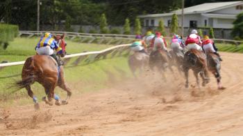
Wounds to the body of the horse can be very large and may initially seem quite severe. Because of the natural tendencies of this prey animal to run from possible danger first and to be concerned about the consequences later, many body injuries result from collisions with trees, fences, wire or other environmental hazards. The horse often is moving quickly when this trauma occurs, and the wounds produced are sometimes superficial and extensive, and they also can be more limited in location but very deep. These deep wounds have the potential to penetrate the abdomen or the chest, and either scenario is a medical emergency.
Advertisement
Wounds to the body of the horse can be very large and may initially seem quite severe. Because of the natural tendencies of this prey animal to run from possible danger first and to be concerned about the consequences later, many body injuries result from collisions with trees, fences, wire or other environmental hazards. The horse often is moving quickly when this trauma occurs, and the wounds produced are sometimes superficial and extensive, and they also can be more limited in location but very deep. These deep wounds have the potential to penetrate the abdomen or the chest, and either scenario is a medical emergency.
The horse often is moving quickly when trauma occurs; though the wounds produced sometimes are superficial and limited in location, they can be extensive and very deep.
Penetrating wounds to the chest have the potential of puncturing lung tissue or damaging the heart. A complete physical examination always should be done in these cases. An evaluation of mucous membrane color, heart rate, respiratory rate and a quick determination of the packed cell volume (PCV) should give the clinician a rapid assessment of possible lung puncture and/or internal bleeding. Crepitus, or air under the skin, also should be looked for because this might indicate some loss of integrity to the respiratory system. Ultrasound of the chest, done between the rib spaces, may also provide some useful information and free air or fluid may be appreciated.
The skin flaps produced by this type of trauma can be impressive, but good cleaning, attention to closure by layer, good apposition and drain placement, if necessary, usually results in good cosmetic results.
Penetration into the abdomen will result in peritonitis at best and possible intestine or organ rupture as a worst-case scenario. Exploration of the penetrating wound is necessary in all cases, and abdominal wound evaluation also can benefit from ultrasound examination, rectal examination and possible abdominocentesis. The presence of bacteria, ingesta and/or blood following an abdominal tap post body trauma is a good indicator that the wound suffered has compromised the abdomen.
Horses with either chest or abdominal trauma should be started on intravenous broad-spectrum antibiotics, and fluids might be necessary to stabilize the horse if bleeding is noted. Referral and transfer to a surgical clinic may be necessary in these cases and a chest/abdominal bandage should be applied once the wound area has been cleaned and stabilized.
Variable blood supply to the lower leg, lack of muscle, fat and extra skin tissue make these wounds very difficult to deal with, but primary closure within the four-hour "golden period" allows for the best results.
Occasionally, horses will be impaled while jumping over obstacles. These boards or poles may have to be left in the horse until the animal can arrive at a surgical facility where aggressive emergency care can be given as the foreign object is removed. If the board or pole is removed in the field, special care and attention should be given to ensure that all pieces are recovered as the splinters can be small and difficult to locate.
Lacerations and punctures to the lower leg in areas over joints or tendons are a combination of superficial injuries to the skin and potentially serious damage to deeper structures.
Advertisement
The majority of body wounds that do not involve deep penetration are usually more sensational than serious. The skin flaps produced by this type of trauma can be impressive, but good cleaning, attention to closure by layer, good apposition and drain placement if necessary usually results in good cosmetic results. Multiple areas of subcutaneous sutures help hold large flaps in place as they heal. Penrose drains or other drain types should be placed in the ventral areas of large flap wounds or in wounds that may be infected. This allows for flushing of these injuries with saline and antibiotics, and pulls the resultant fluid and bacteria away from the subcutaneous space. Even if the skin margins of some parts of the wound are traumatized and may not be viable, it is still advisable to use that tissue initially to promote a granulation response in the deeper part of the wound. Resist the temptation to cut away flaps or strips of muscle or skin because any questionable tissue always can be removed at a later date and may survive better than first thought.
Lower-leg injuries that may or may not involve joints, ligaments and other structures must be monitored closely. Attention improves healing significantly and ultimately generates more practice income.
If motion causes undue stress on the suture or staple line, then these horses will have to be kept in a restricted area and hand-walked. Some exercise is crucial to improving blood flow and to reducing the chances for adhesions.
Leg wounds
Lacerations and wounds to the lower leg of the horse are perhaps the most common equine injuries. Trauma is the most common cause and includes fence injuries (board and wire), kicks, and other environmentally caused wounds. Because of the variable blood supply to the lower leg of the horse and because of the lack of muscle, fat and extra skin tissue, these wounds can be very difficult to deal with. Primary closure within the four-hour "golden period" allows for the best results, but these wounds often occur at pasture, and they are not discovered until much later. Meticulous cleaning, scrubbing and flushing of these wounds are required, and it is not unusual for the wound-preparation time to equal or exceed the time taken to suture or staple the injury.
Post operatively, a well-fitted supportive leg wrap should be applied. The use of an absorptive non-stick pad next to the sutured area can help with initial healing. Feminine pads and liners work very well in this capacity. Antibiotics may be given via regional perfusion if contamination/infection is suspected.
Occasionally, sutured lower-leg wounds are under great tissue stress. Wounds across a flexion/extension surface, such as across the front of the fetlock or down the back of the pastern, may be under tension as a horse moves. These repairs have little chance of success as the constant motion weakens the suture line during time. Wounds of this nature may benefit from the placement of a cast. Cast application can be done under general anesthesia or under sedation in the standing horse. Recovery from general anesthesia for a horse wearing a cast is a difficult procedure, so cast placement under standing sedation is used whenever possible. This is especially true if the wound itself was successfully repaired under sedation. There is often little practical reason to subject the horse to the additional stress/risk of general anesthesia.
DVM Newsbreak
Cast management must be constant and vigilant because cast rubs and the resultant tissue trauma can be more devastating than the initial wound. Monitor the cast for heat, irritation and observe the horse for a decrease in use of the leg or for any signs of discomfort. Thermography, the use of a special ultra-sensitive camera to detect changes in heat in the body, has proven to be especially good at monitoring casts. If the camera shows even a small increase in heat in some area of the cast, then studies have shown that those areas will develop sores. Knowing this, the veterinary clinician can make a small partial thickness cut in the cast over the effected spot to relieve the pressure. In this way casts can be maintained longer and healing has a better chance of continuing without the starts and stops of multiple cast changes.
Lacerations and punctures to the lower leg in areas over joints or tendons are a combination of superficial injuries to the skin and potentially serious damage to deeper structures. These injuries are also considered medical emergencies. Joints and tendon sheaths should be evaluated for puncture by tapping into the suspected area from a location opposite to the site of trauma. If a joint tap reveals bacteria or blood, then it is a logical assumption that the trauma has caused penetration into the joint and that an infection is likely. Immediate and aggressive use of antibiotics is necessary along with copious joint lavage or tendon sheath flushing.
Treating equine lower-leg injuries that may or may not involve joints, ligaments and other structures must be monitored closely.
"Be persistent," says Dr. Reed Hanson, surgeon at Auburn University's College of Veterinary Medicine. "Set up specific appointments to return and recheck these wounds rather than relying on clients to evaluate the injuries and report to your office."
DVM Newsbreak
This hands-on, aggressive approach generates more practice income, and it is simply a better way to monitor the progress of these potentially serious wounds.
"It is a win-win situation for you, the client and, most importantly, for the horse," Hanson says.
Consistent monitoring will allow the practitioner to adjust casts and/or leg wraps, trim damaged tissue as the wound heals, or change and adjust antibiotic dosages. Close monitoring of wounds will improve healing significantly.
Editor's Note: In this second part of a three-part series, Dr. Kenneth L. Marcella discusses body wound care with leading practitioners. Open DVM Newsmagazine's March issue for the first installment on head wounds, or visit our Web site at
Dr. Marcella, a 1983 graduate of Cornell University's veterinary college, was a professor of comparative medicine at the University of Virginia. His interests include muscle problems in sport horses, rehabilitation and other performance issues.
Advertisement
Related Content
Advertisement


Latest CME


Advertisement
Advertisement
Trending on dvm360
1
FDA issues emergency use authorization for ivermectin solution to prevent screwworm in horses
2
Every 30 extra minutes under anesthesia raises complication risk in brachycephalic patients, underscoring the importance of peri-anesthetic management
3
From the CVO: I’m off the clock
4
Paws and profits: VetEvolve names first chief veterinary officer, NVA appoints two board members, and more updates
5