

|Articles|October 1, 2008
Initial stabilization of dogs in respiratory distress (Proceedings)
Author(s)Lesley King, MVB, DACVECC, DACVIM
Respiratory changes occur frequently in small animal patients. It is important to recognize, however, that not all changes in respiration are caused by disease of the respiratory system.
Advertisement
Respiratory changes occur frequently in small animal patients. It is important to recognize, however, that not all changes in respiration are caused by disease of the respiratory system. For example, changes in respiratory function are seen frequently in animals with abdominal or neurologic disease. When faced with a patient that has an increased respiratory rate or difficulty breathing, the clinician must determine whether the changes are caused by disease of the respiratory system, or are secondary to disorders of other systems. The tools used to make this determination can be as simple as observation and auscultation, or as complex as pulse oximetry.
Recognition of the dyspneic animal
In normal inspiration the ribs are pulled cranially and laterally by the external intercostal muscles, contraction of the diaphragm creates negative pressure within the chest, and the abdomen moves outwards. With progression of respiratory distress, as the work of inspiration increases there are greatly increased diaphragmatic excursions, and the secondary muscles of respiration are recruited. The secondary muscles of respiration include the scalene muscles which elevate the first two ribs, the sternomastoids which pull the sternum cranially, and the alae nasi which cause flaring of the nostrils. This is seen as an increase in chest wall excursion, greater abdominal component in expiration, breathing through an open mouth, and dilation of the nares.
The pattern of respiration in the dyspneic animal may be rapid and shallow, or slow and deep, depending on the disease process. Animals with restrictive disease (parenchymal disease that stiffens the lung such as pneumonia, fibrosis, or neoplasia; or disease that restricts the expansion of the lung such as pleural fluid or air) breaths with a rapid, shallow pattern. Animals with obstructive disease (such as airway obstruction or narrowing) tends to take much slower, deeper breaths. Respiration that is noisy is described as stertorous or stridorous, and is usually associated with upper airway or laryngeal disease or obstruction. Wheezes may be rarely be heard associated with disease of the smaller airways.
All of the changes described above are normal responses of the respiratory system to an increased respiratory drive. However, as the work of breathing increases, respiratory muscle failure may ensue as the muscles fatigue. Increased work of breathing may be due to airway obstruction, stiff lungs, or pleural disease. The movements that are seen as respiratory muscles fatigue are called "paradoxical" because they oppose the normal expansion of the chest wall. In normal respiration, both the abdomen and chest move in and out together, allowing maximum expansion of the lungs. In paradoxical respiration: (a) The intercostal muscles may collapse inwards with inspiration as they fatigue, and as greater negative pressures are created within the thoracic cavity; (b) The abdomen may collapse inwards during inspiration for the same reason. In expiration the abdomen appears to move outwards due to increased activity of the abdominal muscles as the diaphragm fatigues. This motion is the opposite of that seen with normal respiration.
Animals in severe respiratory distress assume a posture that impinges least on the work of breathing. If distress is severe enough, they will often have a glazed expression, and will refuse to move, eat or drink, concentrating all of their energy on the movement of air into their lungs. These animals often stand or sit, and if exhausted will tend to lie in sternal rather than in lateral recumbency. They will lift and extend their head and neck in order to maximally open the airway. Often they breathe through an open mouth in order to reduce nasal and pharyngeal airway resistance. Abduction of the elbows allows for maximal movement of the chest wall with each breath.
Oxygen supplementation
Oxygen delivery to the tissues must be prioritized in any critical patient. It can be maximized by careful consideration of pulmonary gas exchange, hemoglobin concentration for oxygen transport, and tissue perfusion for delivery of oxygen to the cells. In any respiratory distress situation, definitive therapy is usually accompanied by oxygen supplementation.
Oxygen supplementation may also be very beneficial in situations when the presence of hypoxemia might not be intuitively obvious by observation. Tachypnea may be wrongly attributed to pain when it really is caused by hypoxemia. Dogs that are flat out because of shock or neurologic involvement may be unable to manifest the typical clinical signs of respiratory distress. The observation of pink mucous membranes does not rule out the possibility of clinically significant hypoxemia, since membranes will remain pink until the PaO2 has dropped below 60 mmHg (normal 85-100 mmHg). Similarly, cyanosis may be undetectable in animals with very pale mucous membranes, because insufficient perfusion of the peripheral tissues by blood may preclude the observation of deoxyhemoglobin. The presence of refractory tachycardia or hypotension, ventricular arrhythmias, severe mental depression, or tachypnea should all trigger measurement of oxygenation. The provision of supplemental oxygen is indicated in every emergency trauma or shock situation until it has been confirmed that the animal is stable without it.
Localization and management of respiratory
Upper airway obstruction
- Dyspnea
- Audible stridor or stertor
- Increased inspiratory effort with prolonged inspiratory time
- Change in vocalization
- Exercise intolerance, clinical signs most severe when stressed or exercising
- Excessive panting
- Hyperthermia
Common differential diagnoses of upper airway obstruction
- brachycephalic syndrome
- laryngeal paralysis
- tracheal collapse
- nasopharyngeal polyps (feline)
- aspirated foreign bodies
- upper airway neoplasia
- retropharyngeal masses, abscesses or hematomas
Emergency management of upper airway obstruction
Oxygen supplementation: O2 cage best
Rest, sedation if necessary, minimal stress
- Acepromazine 0.01-0.05 mg/kg IV or IM if cardiovascularly stable
- Anti-inflammatory to immunosuppressive doses of corticosteroids unless contraindicated
- Dexamethasone 0.1-0.5 mg/kg IV or IM
If collapsed or sedated, extend head and neck and pull tongue out of the mouth
Monitor temperature, vigorous efforts to cool if needed
Fluid therapy if dehydrated or hypovolemic
Emergency tracheostomy or intubation if no response to medical management
Lower airway/bronchial disease
- Cough
- Exercise intolerance
- Dyspnea (severe cases, expiratory dyspnea in cats with feline asthma)
- Increased bronchovesicular sounds
- Wheezes
Common differential diagnoses for lower airway disease
- feline asthma
- chronic bronchitis
- neoplasia
- aspirated foreign body
Advertisement
Emergency management of lower airway disease
Oxygen supplementation
Rest, sedation not usually required
Minimal stress
Anti-inflammatory to immunosuppressive doses of corticosteroids
- Dexamethasone 0.1-0.5 mg/kg IV or IM, or
- Prednisone 0.5-1 mg/kg IV, IM, or PO
- Bronchodilators
- Terbutaline 0.01 mg/kg IV or IM, or
- Aminophylline 5.5 mg/kg IV
Cough suppressants
- Butorphanol 0.2-0.4 mg/kg IV or IM, 0.5-1 mg/kg PO, or
- Hydrocodone 1.25-5 mg/kg PO
Antibiotics
Thoracic radiographs to rule out pulmonary alveolar disease
Consider tracheal wash for culture and cytologic examination
Pulmonary parenchymal disease
- Hypoxemia
- Increased respiratory rate or dyspnea
- Restrictive respiratory pattern
- Harsh bronchovesicular sounds
- Crackles
- Nasal discharge
- Cough (often productive)
Common differential diagnoses for pulmonary parenchymal disease
- pneumonia - bacterial, aspiration, viral, fungal, parasitic
- cardiogenic pulmonary edema
- non-cardiogenic pulmonary edema
- chronic fibrosis/end-stage lung disease
- thromboembolic disease
- neoplasia - primary, metastatic
- pulmonary contusions or hemorrhage
- acute lung injury and ARDS
- pulmonary inflammatory disease (eosinophilic or lymphocytic)
Emergency management of dyspnea due to pulmonary parenchymal disease
Oxygen supplementation
Rest and minimal stress
Vascular access
Thoracic radiographs if possible
Medical management according to most likely diagnosis:
Bronchopneumonia:
Tracheal wash for culture and cytology
Stable:
- Enrofloxacin 5-15 mg/kg PO SID or
- Amoxicillin/clavulanate 14-22 mg/kg PO BID
Unstable:
- Enrofloxacin 5-15 mg/kg IV SID and ampicillin 22 mg/kg IV TID or
- Gentamicin 6 mg/kg IV SID and ampicillin 22 mg/kg IV TID or
- Cefotaxime 20-50 mg/kg IV QID or
- Ticarcillin/clavulanate 50 mg/kg IV QID
- Nebulization and coupage
Pulmonary edema:
Electrocardiography
Echocardiography
- Furosemide 0.5-2 mg/kg IV or IM q 6-12h
- Nitroglycerin paste ¼ to 1 inch cutaneously or
- Nitroprusside 2-10 mcg/kg/min
- Dobutamine 5-10 mcg/kg/min
Hemorrhage
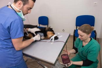
Fresh whole blood transfusions
- Vitamin K 2 mg/kg SQ or PO BID
Pulmonary thromboembolism
Fresh frozen plasma
- Heparin 100-300 U/kg q 6h SQ or 10-50 U/kg/hr IV CRI
Pulmonary inflammatory disorders
- Dexamethasone 0.25-0.5 mg/kg IV or IM or
- Prednisone 0.5-1 mg/kg IV, IM or PO
Pleural space disease
- Increased respiratory rate and effort
- Dyspnea
- Cough
- Dull or diminished lung sounds on auscultation
- Fever
- Weight loss and lethargy
Differentials for pleural space disease
- pyothorax
- non-bacterial exudates
- chylothorax
- hemothorax
- transudates (pure and modified) eg right heart failure
- neoplasia
- lung lobe torsion
- pneumothorax
- diaphragmatic hernia
- pleural neoplasia or mass
Emergency management of pleural space disease
Oxygen supplementation
Rest and minimal stress
Vascular access
Thoracocentesis
Thoracic radiographs if possible after thoracocentesis
Fluid analysis:
- Cell counts
- Cytology
- Aerobic and anaerobic culture
- Biochemical analysis if indicated (triglycerides)
References Available On Request
Advertisement
Related Content
Advertisement


Latest CME


Advertisement
Advertisement