|Articles|February 1, 2003
Histopathology of lesions important for GME diagnosis
Author(s)Johnny D. Hoskins, DVM, PhD, DACVIM
Q: Could you provide a brief review on the causes and management of granulomatous meningoencephalomyelitis (GME) in dogs?
Advertisement
Q: Could you provide a brief review on the causes and management of granulomatous meningoencephalomyelitis (GME) in dogs?
A: Granulomatous meningoencephalitis (GME) occurs primarily inadult female dogs, most often in purebred small breed dogs. Disseminatedgranulomatous lesions primarily affect the white matter of the cerebrum,caudal brain stem, cerebellum and cervical spinal cord and cause multifocalforebrain signs, whereas seizures may be the primary sign if a focal granulomatouslesion is present in the cerebrum.
Animals with the disseminated granulomatous disease often show an acuteand fulminating clinical course, whereas those with focal granulomatousdisease tend to have subtle signs at the beginning and a more prolongedclinical course. GME is a progressive disease.
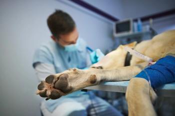
Definitive diagnosis of GME is difficult and involves histopathologicexamination of lesions obtained by way of stereotactic biopsy, open surgicalbiopsy, or post-mortem examination. Ante-mortem diagnosis more commonlyinvolves cerebrospinal fluid analysis, brain/spinal cord imaging with CTor MR imaging, and the elimination of other encephalitides such as causedby canine distemper, Neospora caninum, Toxoplasma gondii and the fungalinfections (blastomycosis, histoplasmosis, coccidioidomycosis, cryptococcosisand aspergillosis) by specific serologic testing procedures. The cerebrospinalfluid collected from a suspected dog typically shows an elevated proteincontent (40-1,000 mg/dl) and increased white blood cell count (primarilyof mononuclear cells, both lymphocytes and macrophages). The most consistentdiagnostic findings from the cerebrospinal fluid analysis are obtained beforeany glucocorticoids are administered.
The cause of GME is still unknown; however, immune-mediated, infectiousand neoplastic causes have been proposed. GME is most likely an immune-mediateddisease of the nervous system.
Glucocorticosteroid and radiation therapy
Symptomatic therapy with glucocorticosteroids has proved successful intemporarily alleviating clinical signs and/or slowing disease progression.
Prednisone has been the most frequently used glucocorticoid; dosagesranging from 2 to 4 mg/kg daily appear to be adequate. Dexamethasone andmethylprednisolone are useful too. The response to steroid therapy variesand cessation of glucocorticoid therapy is invariably associated with rapidand dramatic clinical deterioration, and therefore, glucocorticosteroidsshould not be discontinued.
Advertisement
Long-term, high-dose corticosteroid therapy commonly leads to undesirableside effects such as gastrointestinal ulceration, hepatic dysfunction, andeither iatrogenic hyperadrenocorticism or iatrogenic hypoadrenocorticism.Radiation therapy may prove to be very beneficial for the treatment of focalGME. Dogs treated with radiation therapy for focal GME lesions may havesignificantly longer survival times (median >404 days) than dogs withfocal GME lesions that do not undergo radiation therapy.
The dogs with multifocal GME tend to die either before or during radiation.Radiation therapy is costly and involves daily general anesthesia. Sideeffects of radiation include moist epithelial desquamation, mucositis, keratitis,keratoconjunctivitis and brain necrosis as early effects and blindness,cataracts, and bone necrosis as late effects.
New therapies
Because the long-term side effects of corticosteroids and the expenseand relative unavailability of radiation therapy are less than desirable,new adjunctive and/or alternative medical therapies are preferred. For morespecific details refer to the recent article: Cuddon PA, Coates JR, MurrayM: New Treatments for Granulomatous Meningoencephalomyelitis. Proc 20thAnnual Forum ACVIM 20:319-321, 2002.
Cytosine arabinoside therapy
Cytosine arabinoside (Ara-C) may be used in the treatment of GME in dogs,either as a sole treatment or in conjunction with prednisone. Ara-C, anarabinose-containing nucleoside, crosses the blood-brain barrier in dogsand acts on mitotically active cells by being inserted into DNA moleculesthat result in premature chain termination.
Ara-C has been used to treat CNS neoplasias in dogs and has been usedalone or in combination with other drugs to treat systemic lymphoma andmyeloproliferative disorders. Cytosine arabinoside has also been used intrathecallyto treat CNS lymphoma and appears to effectively reduce the developmentof CNS metastases when administered systemically to cats with renal lymphoma.The reported dosage in dogs for the treatment of neoplasia is variable,ranging from 100 mg/m2 as an intravenous, constant rate infusion, or subcutaneousadministered drug for four days with repeat dosing every three to four weeksup to 600 mg/m2 more than 48 hours and a repeat treatment course in threeto four weeks. Others believe that the drug should be administered at alow-dose constant schedule at 10 mg/m2 daily or twice daily subcutaneously.The most significant side effect of ara-C in dogs is myelosuppression, whichtypically occurs at a nadir of seven to 14 days. Other reported side effectsmay include vomiting, diarrhea and hair loss.
An effective protocol for the long-term treatment of GME in dogs is subcutaneousinjection of ara-C at a dose of 50 mg/m2 twice daily for a period of twoconsecutive days.
This regimen is repeated initially every three weeks. Gloves should beworn when administering this agent. A CBC is performed 10-14 days afterthe first course of cytosine therapy and then periodically throughout thecourse of treatment (usually once every two to three months). It is bestto institute ara-C therapy initially in combination with prednisone (1 mg/kgorally twice daily). After the second round of ara-C injections, a reductionof the prednisone therapy is done by tapering the prednisone.
ara-C may also be used as a sole agent in some dogs with GME. ara-C (FauldingPharmaceuticals, Elizabeth, NJ) is supplied as a 20 mg/ml injectable solutionin 5 ml (100 mg) vials. The cost is about $1.88 per ml.
Procarbazine therapy
Procarbazine is an antineoplastic drug initially synthesized as a potentialmonoamine oxidase (MAO) inhibitor. Procarbazine is lipid-soluble, crossesthe blood-brain barrier and alkylates DNA and affects RNA and protein synthesis.In veterinary oncology, procarbazine is infrequently used as an adjunctivetreatment as part of the MOPP [mechlorethamine, oncovin (vincristine), procarbazine,prednisone] rescue protocol for lymphomas.
Procarbazine has now been used for treatment of GME as an adjunctivetherapy to prednisone or alone. Procarbazine is well absorbed from the gastrointestinaltract and is metabolized by erythrocytes and by microsomal enzymes in theliver to azoprocarbazine. It is widely distributed in the body includingthe cerebrospinal fluid.
The kidney is the major excretion route and less than 5 percent of thedrug is excreted unchanged. Myelosuppression, principally thrombocytopeniaand leukopenia, may be a dose-limiting factor. The nadir of thrombocytopeniais usually about four weeks. Other side effects may include nausea, vomitingand hepatic dysfunction. Procarbazine can sometimes cause neurotoxicityresulting from the passage across the blood-brain barrier but also fromits affects as a monoamine oxidase inhibitor.
Adverse effects may arise with co-administration with a number of drugssuch as ephedrine, isoproterenol, epinephrine, tricyclic antidepressantssuch as imipramine and amitriptyline, narcotics, antihistamines, phenothiazinesand barbiturates. Neurotoxic effects are associated with the peripheraland central nervous systems. Central nervous system effects include bothsedation and agitation. The peripheral effects of procarbazine include lossof tendon reflexes, paresthesia and myalgia. The monoamine oxidase inhibitoreffects cause an increased sensitivity to catecholamines. Procarbazine (matulane)is available as a 50-mg capsule (about $0.70/capsule).
Because of the small size of dogs affected with GME, procarbazine isreformulated into an elixir. It is recommended that a pharmacist re-compoundthe drug because of its toxicity in the powder form. To make a 10 mg/mloil-based solution mix five 50 mg capsules, add some oil-based flavor (liver,chicken, fish) drops, 0.25 tsp silica gel (to keep in suspension), and graduallyadd and mix 25 ml of sesame oil. The expiration date is 30 days. Glovesshould be worn when handling this agent. The oral hydrochloride salt isunstable in aqueous solutions. Procarbazine is administered orally at adose of 25 to 50 mg/m2 daily.
The CBC should be monitored once weekly for the first month, then monthlythereafter. After the first month of therapy, attempt to reduce the frequencyof the dose to every other day.
Newsletter
From exam room tips to practice management insights, get trusted veterinary news delivered straight to your inbox—subscribe to dvm360.
Advertisement
Related Content
Advertisement