

|Articles|August 1, 2009
Degenerative myelopathy – diagnosis and treatment (Proceedings)
Canine degenerative myelopathy is a spontaneously occurring, adult-onset, progressive spinal cord disease.
Advertisement
Canine degenerative myelopathy (DM) is a spontaneously occurring, adult-onset, progressive spinal cord disease. Degenerative myelopathy was first described by Averill as a spinal cord disorder that predominates in German Shepherd dogs. If decreased pelvic limb reflexes are observed, nerve root involvement is presumed and the disease termed chronic degenerative radiculomyelopathy. Initially thought to be specific to the GSD, it also was designated German Shepherd Dog myelopathy. This disease is not uncommon in some pure bred dogs with an overall prevalence rate of 0.19%. Although the German Shepherd Dog is the most commonly affected breed, DM has been reported in other breeds and most recently in the Pembroke Welsh Corgi (PWC). Higher disease prevalence has been determined in a number of other purebred dogs, such as the Boxer, Rhodesian Ridgeback, and Chesapeake Bay Retriever.
Signalment and disease duration
There is no sex predilection. Age of onset of neurologic signs is usually 8 years and older in large breed dogs with DM. A study in PWCs reported a mean age of onset of 11 years. The clinical course of DM can vary up to 3 years after the suspected diagnosis with a mean time for disease duration to be 6 months in larger breeds of dogs (Figure 1). The pet owners often opt for euthanasia when their dogs lose the ability to support weight in the pelvic limbs. Breeds of smaller size allow the pet owner to give the appropriate care for their pet over a longer time. The median time of disease duration in the PWC was 19 months. Longer survival times in DM affected dogs may also be attributed to increased access to physical rehabilitation facilities for companion animals.
Figure 1: Breeds representing mean age at onset, mean age at death and mean duration of clinical signs for Pembroke Welsh Corgi (PWC), Chesapeake Bay Retriever (CBR), Boxer, and Rhodesian Ridgeback (RR) with a diagnosis confirmed by histopathology or presumptive based on normal imaging
Progressive, asymmetric upper motor neuron (UMN) paraparesis and lack of paraspinal pain are key clinical features of DM. Physical examination findings include loss of muscle mass in the caudal trunk muscles, pelvic limb weakness, and worn nails. Descriptions for severity of loss of muscle mass have differed. Most reports attribute this loss to disuse but flaccidity has been noted in the later stage of disease which suggests neurogenic muscle atrophy. Gait deficits at time of onset show spastic paresis and general proprioceptive ataxia (loss of joint position sense as described for domestic animals). Asymmetric weakness at disease onset also is frequently reported. Most large breed dogs progress to nonambulatory paresis or paraplegia within 6 to 12 months from time of diagnosis. If the disease progresses over a longer duration, clinical signs will ascend to affect the thoracic limbs. If euthanasia is delayed, the clinical signs will cause flaccid tetraparesis/plegia and other lower motor neuron (LMN) signs. Due to its smaller size and longer disease duration, this was a more common scenario in the PWC.4 At disease onset, descriptions of spinal reflexes correspond with UMN paresis. Decreased reflexes noted with the patellar and withdrawal reflexes occur in the latter disease stage. Urinary and fecal continence usually are spared until the latter disease stage.
Pathogenesis
Until recently the pathogenesis of canine degenerative myelopathy had remained unknown. Griffiths and Duncan originally hypothesized DM to be a "dying-back disease" confined to the CNS suggesting a toxic etiology. Recently, studies of the brain of DM affected GSDs showed neuronal degeneration and loss in some brainstem nuclei. Johnston et al. suggested that a defect in the neuron, itself that may lead to abnormal axonal transport and degeneration in the distal axon.
An immunologic role in the pathogenesis of GSD DM has been proposed based upon observations of depressed responses to thymus-dependent mitogens and increased concentrations of circulating immune complexes. Although immune-related degenerative disease is a plausible theory, immunosuppressive therapies have shown no long-term benefits in halting the progression of DM. Based on the immunologic hypothesis, Clemmons et al. (FASEB, 2006) claimed that there is a point mutation in the hypervariable region 2 of DLA-DR� and termed allele *1101J and reported homozygosity in DM affected GSDs. A more recent study showed that data did not provide evidence for involvement of DLA-DRBI in DM and indicated that the test offered by the University of Florida was not predictive for DM.
Early DM studies suggested a genetic cause. The first report to suggest familial disease of DM was in the Siberian Husky. Based on frequency of affected dogs having affected relatives, there is clear familial aggregation in the PWC, Rhodesian Ridgeback (Coates – unpublished data), Boxer (Coates – unpublished data), and Chesapeake Bay Retriever (personal communication – Dr. Sam Long, University of Pennsylvania). The lack of pedigree data and histopathologic confirmation in presumed affected dogs of early generations from affected families make it difficult to evaluate the inheritance pattern of DM. Currently, an autosomal recessive inheritance pattern is presumed. Obtaining DNA samples from relatives of DM affected dogs has been challenging due to the late-onset of DM and parents being deceased.
We used the canine SNP chip (Affymetrix v2) to genotype DNA samples from affected and healthy dogs. Genome-wide association mapping analysis revealed a peak with strongest association on canine chromosome 31 which also showed markers that contained the canine the superoxide dismutase (SOD1) gene. Resequencing of SOD1 in normal and affected dogs revealed a G to A transition, resulting in an E40K missense mutation. Homozygosity for the A allele was associated with DM in five common dog breeds (German Shepherd dog, Boxer, Rhodesian Ridgeback, Welsh Corgi, and Chesapeake Bay Retriever). The frequency of the A allele in a separate "other breeds" control group, consisting of samples from dog breeds in which DM was rarely diagnosed, was significantly lower than that for the controls from the affected breeds. Not all animals that are homozygous for this mutation will get the disease. Thus, penetance amongst the mutant homozygotes is incomplete. To summarize, we have discovered a risk factor for dogs to develop DM. We believe that there are other genetic modifiers and environmental factors that could influence whether these dogs at risk actually get DM.
Mutations in the SOD1 gene are known to cause amyotrophic lateral sclerosis (ALS) in humans; also known as Lou Gehrig's disease. The disease derives its name from the combined degeneration of upper and lower motor neurons projecting from the brain and spinal cord. The Greek derivation of amyotrophy means, "muscles without nourishment." Lateral is the location within the spinal cord of axonal disease and sclerosis refers to diseased axons being replaced by sclerosis or "scar" tissue. The disease affects people in their fourth or fifth decade of life. Most people die within 5 years after disease onset.
There are several different forms of ALS in people that lead to progressive paralysis and subsequent respiratory failure. The major hallmarks of criteria for a diagnosis of ALS include signs of upper (brain or spinal cord) and lower (nerve and muscle) motor neuron degeneration, and progressive spread of these signs within a region or to other regions. Degenerative myelopathy affected dogs initially manifest UMN signs, similar to UMN-onset ALS, that progress to also involve the thoracic limbs and later manifest LMN signs. DM affected dogs also have an absence of electrophysiological or neuroimaging evidence of other disease processes that could explain the combination of progressive upper and lower motor neuron dysfunction. Because this is a spontaneously occurring disease and because we consider the dog nervous system more like human than either the rat or mouse, we hope that this model of ALS may be more predictive of outcome in human ALS clinical trials. Mouse and rat ALS models have thus far been disappointing since many drugs that were successful in the mouse/rat models subsequently failed in human ALS. Furthermore, the SOD1 mutation in dogs may offer significant advantages for some studies of ALS pathogenesis.
Diagnosis
Tentative antemortem diagnosis presently is based upon ruling out other diseases causing progressive myelopathy. Common differentials include orthopaedic disease, intervertebral disc degeneration, inflammatory disorders, and spinal cord neoplasia. Neurodiagnostic techniques for evaluation of spinal cord disease include CSF analysis, electrodiagnostic testing, myelography, computed tomography (CT) and magnetic resonance imaging (MRI). A presumptive diagnosis of DM often is made based on lack of clinically relevant compressive myelopathy as determined by myelography, CT/myelography or MRI. Electrodiagnostic testing is used to rule out other disorders causing neuromuscular weakness. Electromyography and nerve conduction studies can be abnormal on DM affected dogs during the latter disease stage. We have confirmed this on nerve and muscle biopsies. Studies specific to the peripheral nervous system in DM affected dogs still need to be pursued. We are using a combination of genetic testing (see below) and neurodiagnostic testing to assist with a presumptive diagnosis of DM.
Definitive diagnosis of DM is determined postmortem by histopathologic examination of the spinal cord. Spinal cord pathology of DM is consistent with a noninflammatory axonal degeneration. Histopathologic studies of DM have been more frequently described in the GSD. Affected dogs have characteristic patterns of axon cylinder vacuolization and drop out in this form of degenerative myelopathy. Lesion distribution involves the spinal cord myelin and axons in all parts of the spinal cord white matter, but affects the mid- to caudal thoracic region most extensively. A recent description of DM in the Pembroke Welsh Corgi characterized the overall distribution of white matter lesions to be similar to that observed in DM of large breed dogs. Previous studies, however, have consistently reported longitudinally discontinuous lesions in affected tracts with a tendency for increased lesion severity within the dorsal portion of the lateral white matter and dorsal white matter. An important histopathologic difference in PWC dogs is the longitudinally continuous nature of lesions within more clearly defined areas in the white matter of the spinal cord. Ascending spinocerebellar and descending rubrospinal and lateral corticospinal tract involvement was especially evident in all spinal cord segments examined. The more severe and well-demarcated lesions in PWC dogs simply may reflect disease longevity. We have additionally documented neuromuscular pathology as evidence by denervation atrophy in muscle, and nerve fiber loss, axonal degeneration and demyelination of myelinated fibers in peripheral nerves.
Treatment and prognosis
Advertisement
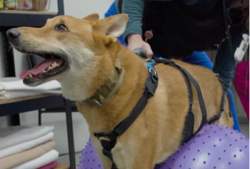
Treatment regimens have been empirical with lack of evidence based medicine. Aminocaproic acid, an antiprotease agent, has been advocated for long-term management of DM; however, there have been no published clinical data to support drug efficacy. While vitamin deficiencies can cause spinal cord degeneration in some species, therapy with parenteral cobalamin or oral tocopherol did not affect neurologic progression in a study of DM affected dogs; moreover, serum concentrations of alpha-tocopheral in DM affected GSDs did not yield significant differences when compared to unaffected DM dogs. Combination therapies with an exercise regimen have been advocated for treatment of DM. Kathmann et al. reported survival data in 22 DM affected dogs that received varying degrees of physiotherapy. Results suggested dogs that received intensive physiotherapy had a significantly longer survival time compared to dogs that received moderate or no physiotherapy. Additionally, dogs that received physiotherapy remained ambulatory longer than those that did not. However, the long term prognosis still remains poor.
Genetic testing
A genetic test is currently available through the Orthopedic Foundation for Animals (OFFA) in association with the Animal Molecular Genetic Diseases Laboratory of the University of Missouri. (
Interpreting results
Normal (n/n)
This dog is homozygous N/N, with two normal copies of the gene. In the seven breeds studied at the University of Missouri in depth so far, dogs with test results of N/N (Normal) have never been confirmed to have DM. This dog can only transmit the normal gene to its offspring, and it is unlikely that this dog or its offspring will ever develop DM.
Carrier (a/n)
This dog is heterozygous A/N, with one mutated copy of the gene and one normal copy of the gene, and is classified as a carrier. In the seven breeds studied at the University of Missouri in depth so far, dogs with test results of A/N have never been confirmed to have DM. While it is highly unlikely this dog will ever develop DM, this dog can transmit either the normal gene or the mutated gene to its offspring.
At-risk (a/a)
This dog is homozygous A/A, with two mutated copies of the gene, and is at risk for developing Degenerative Myelopathy (DM). The research has shown that all dogs in the research study with confirmed DM have had A/A DNA test results, however, not all dogs testing as A/A have shown clinical signs of DM. DM is typically a late onset disease, and dogs testing as A/A that are clinically normal may still begin to show signs of the disease as they age. Some dogs testing A/A did not begin to show clinical signs of DM until they were 14 years of age. Research is ongoing to estimate what percentage of dogs testing as A/A will develop DM within their lifespan. At this point, the mutation can only be interpreted as being at risk of developing DM within the animal's life. For dogs showing clinical signs with a presumptive diagnosis of DM, affected (A/A) test results can be used as an additional tool to aid in the diagnosis of DM. Dogs testing Affected (A/A) can only pass the mutated gene on to their offspring.
Guidelines for breeding
Owners with dogs testing as Carriers (A/N), or At-Risk (A/A) are strongly encouraged to share these results with their attending veterinarian and seek genetic counseling when making breeding decisions. The "A" (mutated) allele appears to be very common in some breeds. In these breeds, an overly aggressive breeding program to eliminate dogs testing A/A or A/N might be devastating to the breed as a whole because it would eliminate a large fraction of the high quality dogs that would otherwise contribute desirable qualities to the breed. Nonetheless, DM should be taken seriously. It is a fatal disease with devastating consequences for the dog, and can be a trying experience for the owners that care for them. A realistic approach when considering which dogs to select for breeding would be to treat the test results as one would treat any other undesirable trait or fault. Dogs testing At-Risk (A/A) should be considered to have a more serious fault than those testing as Carriers (A/N). Incorporating this information into their selection criteria, breeders can then proceed as conscientious breeders have always done: make their breeding selections based on all the dog's strengths and all the dog's faults. Using this approach and factoring the DM test results into the breeding decisions should reduce the prevalence of DM in the subsequent generations while continuing to maintain and improve upon positive, sought after traits.
The allelic frequency may be as high as 75% in breeds prone to DM. (unpublished data, Dr. Gary S. Johnson – Animal Molecular Genetics Laboratory, University of Missouri). We recommend that breeders take into consideration the DM test results as they plan their breeding programs; however, they should not over-emphasize the test results. Instead, the test result should be one factor among many in a balanced breeding program.
References
1. Averill DR. J Am Vet Med Assoc 1978;162:1045.
2. Griffiths IR, et.al. J Small Anim Pract 1975;16:461.
3. Braund KG, et.al. Am J Vet Res 39:1309.
4. Coates JR, et.al. J Vet Int Med 2007;21:1323.
5. Matthews NS, et.al. J Am Vet Med Assoc 1985;186:1213.
6. Kathmann I, et.al. J Vet Int Med 2006;20:927.
7. Johnston PEJ, et.al. Vet Rec 2000;146:629.
8. Waxman FJ, et.al. J Immunol 1980;124:1209.
9. Waxman FJ, et.al. J Immunol 1980;124:1216.
10. Clark LA, et al. Anim Genet 2008;1.
11. Bichsel P, et.al. J Am Vet Med Assoc 1983;183:998.
12. Sutter NB, et.al. Genome Res 2004;12:2388.
13. Lindblad-Toh K, et.al. Nature 2005;438:803.
14. Karlsson EK, et.al. Nature Genetics 2007;39(11):1321.
15. Awano T, et al. Proceed Nat Acad Sci 2009;106 (8):2794-2799.
16. March PA, et al. Vet Path 2009;46:241-250.
17. Johnston PEJ, et.al. Vet Rec 2001;148:403.
18. Previously presented at the 2009 Annual Forum of the American College of Veterinary Internal Medicine in Montreal Canada June 3-6, 2009.
Advertisement
Related Content
Advertisement


Latest CME


Advertisement
Advertisement
Trending on dvm360
1
PerioVive: Regenerative Support Through Hyaluronic Acid for Every Dental Procedure
2
Weekly Vet Report: First clinical use of TMJ arthroscopy in dogs, Lyme-lepto combo vaccine approval, & more
3
Investigational drug shows promise for treating feline obesity
4
New World screwworm: The flesh-eating parasite returning to the spotlight
5