

|Articles|April 1, 2011
Common endocrine dermatopathies in dogs
A look at dermatologic disorders associated with endocrine disease.
Advertisement
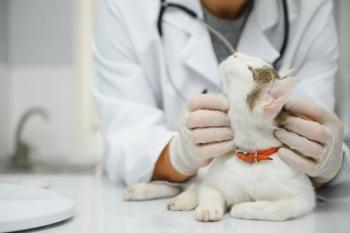
Sam is a 10-year-old 65-lb neutered male black Labrador retriever mix with a history of pruritus, regional hair loss, malodor and a dull coat.
The medical team, using a medical history form (Table 1), determined that Sam has been pruritic for the past six months, and his scratching has become more obvious during the past two months. According to the owners, no other pet or person in the household is affected. Sam has been receiving antibiotics, antihistamines and corticosteroids during the past six months; each has had limited success in resolving his clinical signs.
Table 1: Elements of allergic disease vs. endocrine disease
Sam spends most of his time indoors and eats only a commercial dry adult food. The owners report no fleas in the household. When asked, the owners say Sam's energy level is a little less than normal, and he does not want to play as much as he used to. They have attributed this to his "getting older."
Physical examination shows Sam is mildly pruritic with generalized hair loss that is most noticeable over the back and tail (Photo 1). There's also increased pigmentation on the patient's ventrum; generalized malodor is also evident.
Photo 1: Sam at presentation. Note the generalized hair loss that is most noticeable over the dog's back and tail. (Photos courtesy of Kim Coyner, DVM, Dipl. ACVD, Dermatology Clinic for Animals, Las Vegas, N.V. )
The medical team performs a standard dermatologic diagnostic workup consisting of skin scraping, skin cytology and a dermatophyte culture. Review of the samples shows no parasites on the skin scrape, but numerous Malassezia species and coccoid bacteria are evident on cytology (Photo 2). A thorough initial evaluation shows staphylococcal pyoderma and Malassezia dermatitis.
Photo 2: Malassezia (arrows) and coccoid bacteria on skin cytology.
A three-week antibacterial and antifungal regimen in initiated, and Sam is rechecked in three weeks. During this time, the dermatophyte culture is monitored daily and determined to be negative. The owners report the pruritus and odor have decreased; however, the alopecia has not changed and, in fact, may be worse.
Now the veterinarian must determine the underlying cause of the recently cleared cutaneous infections. Given the information available, an endocrinopathy or an allergy (e.g., atopy, food or parasite hypersensitivity) should be considered. The three most common endocrine disorders to affect a dog's skin are hypothyroidism, hyperadrenocorticism (Cushing's disease) and the more all-inclusive condition known as alopecia X. Several rare endocrinopathies area also known to affect the skin, including estrogen- and testosterone-producing tumors, resulting in abnormal sex hormone concentrations.
Findings suggest Sam has more underlying disorders than a typical allergic patient. Several historical and physical clues suggest endocrine disease. It's important to accurately identify the specific endocrinopathy, since many of them eventually may lead to systemic abnormalities. Since hypothyroidism and hyperadrenocorticism have the most potential for leading to clinical abnormalities and systemic effects, detection of these diseases are the main focus of this article. The following five-step protocol is suggested to help identify dermatologic patients with an endocrinopathy.
1. Identify the signalment
Signalment is only slightly helpful in separating an allergic patient from one with an endocrinopathy. In general, patients presenting with an endocrinopathy are older than the typical allergic patient. Purebred and several breeds of dogs are more likely than others to develop both allergies and endocrinopathies.
Although any animal may be affected, breeds more likely to develop hypothyroidism include golden retrievers, Doberman pinschers, dachshunds, Irish setters, Labrador retrievers, miniature schnauzers, Great Danes, poodles, cocker spaniels, shelties and boxers.
Dogs most prone to hyperadrenocorticism include poodles, dachshunds, German shepherds, beagles, Boston terriers and other terriers.
2. Determine the chief complaint
In general, a dog with an endocrinopathy will be nonpruritic except when the skin is infected. Metabolic changes are common, especially in hypothyroidism, and owners may complain that the pet has decreased energy, increased weakness or acts old. Body shape and conformation may change with weight gain, fat deposition and the development of a distended abdomen. The owner will often mistakenly attribute these changes to normal aging.
3. Get the medical history
The medical team should focus on key elements that may suggest a more severe systemic disease. Some key questions to ask the client include:
- How is the pet's energy level? Are you aware of changes such as lethargy or dull or sluggish behavior by the pet? These changes can be subtle and progressive, and the owner may not volunteer this information until asked specifically.
- Is there an overall problem with or change in the animal's weight? Both hypothyroidism and hyperadrenocorticism can cause weight gain. Hypothyroid animals have a lower metabolic rate, so weight may increase even without an increase in calorie intake. Cushingoid patients may have a pendulous abdomen that can make the pet appear fat.
- Has the animal's coat changed? Hypothyroid dogs are incorrectly assumed to shed excessively. In fact most hairs remain in the telogen phase and, in essence, stop growing. Over time, the coat can appear dull, bleached out and worn, especially at sites of friction created while the animal lies down, sits or wears a collar or harness. The alopecia may be more dramatic or complete on the trunk with hyperadrenocorticism.
- Does the pet have a history of ear infections? Hypothyroid animals can have chronic sustained ear infections, secondary to alterations in keratinization, sebum formation and cutaneous immunity. Remember, though, that most patients with otitis are allergic and not suffering from an endocrinopathy.
- Has there been an increase in water consumption (polydipsia) or urination (polyuria)? With hyperadrenocorticism, there can be a sudden or gradual increase in both.
4. Perform a dermatologic evaluation
Most patients with an endocrinopathy won't show signs of inflammation or erythema of the skin; instead, it may appear cool, especially with hypothyroidism. The trunk is often the site most notably affected, with a symmetrical alopecia of the dorsal trunk or tail (Photo 3A). However, large dogs are known to have alopecia develop at points of wear, including their extremities. Common notable skin changes can include:
Photo 3A: A patient with loss of hair over its tail ("rat tail"), suggestive of an endocrinopathy.
• Symmetrical truncal and tail hair loss, with normal coat on the head and distal legs
• Retention of the fuzzy "puppy coat"
• Hyperpigmentation (Photo 3B)
Photo 3B: A patient with hyperpigmentation of the ventrum, suggestive of an endocrinopathy.
• Excessive shedding or easily epilated hair (Photo 3C)
Photo 3C: A patient with increased regions of hair loss and skin thickening (lichenification), suggestive of an endocrinopathy.
• Cool, thin skin that bruises easily
• Myxedema that affects facial skin folds resulting in a "tragic" expression (Photos 3D and 3E),
Photo 3D and 3E: Patients with excessive thickening of the facial folds ("tragic" expression), suggestive of an endocrinopathy.
• Recurrent or chronic skin infection (e.g., bacterial, Demodex species or dermatophyte)
• Variable coat color changes—notably, bleaching due to slower hair cycle and slower turnover of hair
• Nonpruritic skin (unless there's a secondary infection evident)
• Alopecia on the extremities or distal limbs of larger dogs, especially at the sites of wear or friction
• Thin or atrophic skin
• Comedone formation
• Hard erythematous papules and plaques from calcinosis cutis
• Circumanal gland hyperplasia.
5. Perform clinical diagnostic testing
The time to evaluate the patient for an endocrine disease will depend on the patient's initial history and physical examination findings. In many cases, the veterinarian will address the secondary skin infections that may be causing pruritus at the initial examination and then re-evaluate the patient 21 days after such therapy is finished. If the patient's pruritus and lesions have resolved, but clinical signs are still present (suggesting an endocrinopathy), then screening for hypothyroidism and Cushing's disease is indicated.
Initial screening tests for suspected endocrinopathy include a complete blood count, serum chemistry profile, urinalysis and thyroid screening, including thyroxine (T4), free T4 by equilibrium dialysis and thyroid-stimulating hormone (TSH) measurement. The following changes can be detected in patients with an endocrinopathy:
- Marked serum lipemia. In many patients, their ability to metabolize fat is affected by endocrine disease. In some cases, patients will present with a marked serum lipemia, evident once the blood tube is spun down. Lipemia can occur if a patient's blood is drawn immediately after eating or with other medical conditions. However, if lipemia is noted, the staff should bring it to the veterinarian's attention.
- Mild changes in blood studies. By themselves, mild changes are not indicative of endocrine disease, but they should be reviewed in conjunction with the entire medical database. Hypothyroid patients can present with normocytic, normochromic, nonregenerative anemia. Patients with Cushing's disease often have neutrophilia, monocytosis, lymphopenia and eosinopenia.
- Changes in clinical chemistry. Just as the complete blood count results can reveal changes indicating endocrine disease, the serum chemistry profile findings may show changes, raising the level of suspicion that an endocrinopathy exists. For example, in hypothyroidism, increases in serum cholesterol and lipid concentrations can be observed secondary to improper metabolism of dietary fats. Thyroid hormone is necessary for all facets of lipid metabolism, but it is especially important in the mobilization and degradation of lipids. Liver enzyme activities, such as alkaline phosphatase, aspartate aminotransferase and alanine aminotransferase, may be increased secondary to fatty liver changes in the patient.
Meanwhile, increased alkaline phosphatase is the most common biochemical abnormality in dogs with hyperadrenocorticism, with increased activities being present in 90 percent to 95 percent of these patients. Also an increased cholesterol concentration is seen in 90 percent of dogs with this disorder due to glucocorticoid stimulation of lipolysis.
Glucocorticoids will increase blood glucose and decrease peripheral utilization. Therefore, patients with hyperadrenocorticism can have slight to moderate increases in blood glucose concentration. Lastly, increased alanine aminotransferase activity is likely due to hepatocytes swollen with glycogen accumulation.
- Urinalysis. Patients with hyperadrenocorticism often have dilute urine (urine specific gravity < 1.015). A urine sample also can be used to screen for diabetes mellitus, proteinuria and urinary tract infection, all of which are common sequelae of hyperadrenocorticism.
- Thyroid testing. No one thyroid test is 100 percent diagnostic for hypothyroidism. Basal measurements of total T4, free T4, free T4 by equilibrium dialysis (which is more accurate than free T4) and TSH are the mainstays of testing. Typically, hypothyroid patients have decreased total T4 and free T4 concentrations and increased TSH concentrations. Many factors, such as concurrent illness and drug therapy, will influence the resting total T4 concentration. Even the values set by the laboratory will determine if dogs are hypothyroid; if a reference laboratory sets the bottom of the range too high, normal dogs will appear to be hypothyroid.
Patients with other underlying or concurrent illnesses may be incorrectly determined to be hypothyroid due to a condition called euthyroid sick syndrome. In this syndrome, chronic disease and drug therapy can decrease serum thyroid hormone concentrations, decrease thyroid gland responsiveness to TSH and decrease pituitary secretion of TSH. Once these patients are treated for the primary disease, their thyroid function returns to normal. Disorders known to cause euthyroid sick syndrome include chronic infection (especially deep pyoderma) and hyperadrenocorticism. This can result in a confusing and challenging situation, and the veterinarian will need to discern if the hypothyroidism caused the deep pyoderma, or if the deep pyoderma caused the euthyroid sick syndrome. Drugs that may decrease thyroid hormone concentrations include glucocorticoids, phenobarbital, nonsteroidal anti-inflammatory drugs, penicillin antibiotics, imidazole antifungals, furosemide, diazepam, antineoplastic agents and phenothiazines.
- Adrenal cortisol testing. Although multiple clinical diagnostic tests are available, there isn't always a single correct diagnostic pathway that will point to Cushing's disease. Some clinical tests that can help in diagnosis include:
—Urine cortisol/creatinine ratio. In animals with Cushing's disease, excess cortisol is excreted by the kidneys, increasing the amount of cortisol in comparison to the normal concentrations of creatinine in the urine.
—ACTH stimulation. In a dog fasted 12 hours, serum is obtained to determine a pre-stimulation cortisol concentration. Then ACTH (pituitary hormone) is given, either intramuscularly or intravenously, depending on the form of ACTH. One or two hours later (again, depending on the ACTH form), a post-stimulation cortisol concentration is obtained. Hyperadrenocortical patients show a significant increase (far above normal response to ACTH) of cortisol concentrations after stimulation due to hypertrophic adrenal glands. Although this test can confirm a patient suffers from hyperadrenocorticism, it does not distinguish the pituitary-dependent from the adrenal-dependent (adrenal tumor) form of Cushing's disease.
—Low-dose dexamethasone suppression. In a fasted animal, serum is obtained for measurement of cortisol at the time of injection of dexamethasone sodium phosphate and then again four and eight hours later. An intravenously administered dexamethasone dose of 0.015 mg/kg is used. A patient with a normal pituitary-adrenal axis will respond by suppressing the endogenous cortisol to concentrations below the reference range (different with each laboratory) at both four and eight hours post-injection. A patient with hyperadrenocorticism won't respond to the dexamethasone; subsequently the endogenous cortisol concentration will remain above the reference range at the eight-hour measurement. The four-hour measurement can be useful because some patients with pituitary-dependent hyperadrenocorticism will have suppressed concentrations of cortisol at four hours, but the concentration will "escape" or rise above the normal range at the eight-hour measurement. In this instance, the test will confirm both the disease (Cushing's) and the form (pituitary-dependent). Note, some pituitary-dependent cases of Cushing's disease will not show a suppressed cortisol concentration at four hours post-dexamethasone. It's expected that a patient with a functional adrenal tumor will have endogenous cortisol concentrations that remain increased, or not suppressed, at both four and eight hours post-dexamethasone. This test is considered by many to be more sensitive at confirming a patient with Cushing's, but not as specific as an ACTH stimulation test. Other systemic illnesses, such as diabetes mellitus or renal or liver disorders, may result in false-positive low-dose dexamethasone suppression.
—High-dose dexamethasone suppression. This test can be performed to distinguish the pituitary-dependent from the adrenal-dependent form of hyperadrenocorticism, but only after confirming this disease exists by either the ACTH stimulation test or the low-dose dexamethasone suppression test. In this test, a high-dose (1 mg/kg) of dexamethasone is injected intravenously. With pituitary-dependent Cushing's disease, most pituitary tumors will decrease the ACTH concentration in the presence of higher dexamethasone concentrations. The decreased ACTH decreases endogenous cortisol concentrations. In adrenal-dependent Cushing's disease, three out of four adrenal tumors won't suppress cortisol concentrations after the injection of the high-dose dexamethasone. Note, both false positive and false negative results are possible when using high-dose dexamethasone to differentiate pituitary from adrenal forms of hyperadrenocorticism.
—Abdominal ultrasonography. Once hyperadrenocorticism is suspected through routine clinical diagnostic tests or a Cushing's screening test (i.e., urine cortisol/creatinine ratio), abdominal ultrasonography can detect adrenal gland changes. Specifically, ultrasonography can help detect three disorders: 1) pituitary-dependent disease, or enlargement (one to two times normal size) of both adrenal glands due to increased ACTH production from the pituitary gland, resulting in bilateral hyperplasia; 2) adrenal tumors, that is, moderate enlargement of one adrenal gland and atrophy of the second gland secondary to negative feedback of increasing cortisol concentrations and decreasing ACTH concentrations; and 3) pheochromocytoma (epinephrine-producing tumor of the adrenal medulla), that is, a moderate-to-severe enlargement of one adrenal gland, while the other gland usually is normal in size.
Summary
Identification and diagnosis of endocrinopathies requires close attention to medical history, physical examination findings, identification of secondary infections and a thorough clinical diagnostic database. The medical team must be able to isolate abnormal findings that may suggest the presence of a subtle underlying disease process. These changes help the veterinarian determine if more specific endocrine or allergy testing should be done.
Dr. Lewis sees dermatology patients in California, Arizona, Nebraska, New Mexico, Nevada and Utah. In 1991, he established Dermatology for Animals, PC. Dr. Rosenfeld is the founder and president of VTEC. He has practiced small-animal critical care and emergency medicine for 20 years and is currently working as a general practitioner and running a mobile ultrasound practice in Cape Cod, Mass.
SUGGESTED READING
1. Scott DW, Miller Jr. WH, Griffin CE. Muller and Kirk's small animal dermatology. Baltimore, Md: Elsevier, 2000.
2. Feldman EC, Nelson RW. Canine and feline endocrinology and reproduction. 3rd ed. Baltimore, Md: Elsevier, 2003.
3. Rosenfeld A. The veterinary medical team handbook. Ames, Iowa: Wiley Blackwell, 2007.
Advertisement
Related Content
Advertisement


Latest CME


Advertisement
Advertisement
Trending on dvm360
1
Weekly Vet Report: First clinical use of TMJ arthroscopy in dogs, Lyme-lepto combo vaccine approval, & more
2
New World screwworm: The flesh-eating parasite returning to the spotlight
3
Veterinary hospice offers cats and clients familiar comforts (Part 1): Familiarize clients with veterinary hospice
4
Survey highlights gaps in canine pruritus management expectations
5