

|Articles|August 1, 2009
Cardiocerebral resuscitation (Proceedings)
Author(s)Mary Tefend Campbell, CVT, VTS (ECC)
Predisposing factors that may preclude a cardiocerebral arrest should be eliminated when possible for every critical patient.
Advertisement
Predisposing factors that may preclude a cardiocerebral arrest should be eliminated when possible for every critical patient. For example, electrolyte and acid-base imbalances should be identified and corrected, patients with respiratory distress should be intensely observed and receive supplemental oxygen. Shock, trauma, sepsis, hypothermia, and cardiac arrhythmias may all result in a cardiocerebral arrest if not treated aggressively and accurately. In anesthetized patients, anesthetic overdose, hypoventilation, inadequate fluid volume, and extreme hypothermia should be avoided, as these factors may predispose a patient to an arrest. Positive resuscitation statistics are highest on those patients that were healthy before and whose arrest was initiated by acute and easily correctable problems
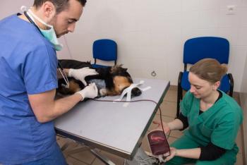
Cardiopulmonary arrest (CPA) is the cessation of functional ventilation and effective circulation. Apneustic gasps may occur, but this action does not produce effective ventilation. Similarly, cardiac electrical or muscle activity may persist, but effective tissue perfusion does not occur. Clinical signs of CPA include absence of auscultable heartbeat, lack of palpable pulse, apnea or agonal gasping, absence of bleeding, loss of consciousness, and pupillary dilation.
The patients most likely to be successfully resuscitated are those which were normal prior to the incident resulting in an arrest, as well as those in which resuscitation techniques were initiated immediately. It is important to have a "CPCR station" at the hospital which is kept in a state of continual readiness. A crash cart should contain an assortment of endotracheal tubes, catheters, syringes, gauze, and cardiac drugs already drawn up in appropriate sized syringes. In addition, sterile instruments should be available. The station must be near an oxygen source, an electrocardiogram, and a defibrillator with internal and external paddles. It is often helpful to have an emergency chart with a list of cardiac drugs and dosages posted in this area.
When an arrest occurs, the veterinary team must respond with urgency, but not with panic. Proceed in an orderly fashion though the "ABC's" of CPR, keeping in mind not to jump out of sequence.
"A" is for airway
The first step of CPR may very well be the most important, since subsequent efforts are futile if the animal is not receiving oxygen. As soon as the clinician has determined that CPA has occurred (no palpable pulse, no auscultable heartbeat), the animal should be intubated. Due to the importance of this step, the clinician must make SURE that the endotracheal tube is in the trachea. This can be done by 1) direct visualization of the tube entering the glottis or 2) palpation of the tube in proper position between the arytenoid cartilages, or 3) confirmation via capnography. If the tube is correctly placed within the trachea, upon ventilation, a number should be greater than zero. The cuff should be inflated and the tube secured in place, with a visible marker to monitor for tube migration. If the airway is not patent and intubation is not feasible, an emergency tracheostomy should be performed immediately.
"B" is for breathing
Once the airway is established, the animal should be ventilated several times with 100% oxygen and then quickly checked for return of the heartbeat by either auscultation or palpation of femoral pulses. If the patient is in surgery, the inhalant anesthetic should be turned off immediately; if on an anesthetic ventilator, increase the FiO2 to 100% with the tubing and bag flushed or replaced. Artificial ventilation is administered by intermittent positive pressure breathing using either an Ambu-bag with 100% oxygen or an anesthetic machine. Animals should be hyperventilated, with rapid ventilations occurring simultaneously with cardiac compressions. Simultaneous ventilations and external chest compressions will increased blood flow and cardiac output due to the effect of generalized increased intrathoracic pressure on the thoracic pump mechanism. The increased ventilatory rate tends to thwart probable metabolic acidosis by removing excess CO2 that invariably develops with CPA.
Recommended peak inspiratory pressures are 20 cm water (dog) and 15 cm water (cat). When ventilations are simultaneous with compressions, higher pressures (20-30 cm water) may be required. Animals with pneumothorax, pleural fluid, pulmonary edema, hemorrhage, or pneumonia may also require higher pressures to obtain lung expansion. Inspiratory time should be less than 1.5 seconds to allow for adequate venous return as the intrathoracic pressure drops. It is also important that expiratory pressure be allowed to fall to 0 cm H2O between forced inspirations. Generally, positive-end-expiratory-pressure (PEEP) is not indicated in CPA because it diminishes venous return, cardiac output, and the effectiveness of the thoracic pump mechanism. Airway tubing should be monitored for pulmonary fluid which may impede ventilation; suction should be utilized to remove fluid, in addition to removing tubing temporarily to tip the patient to remove fluid from the trach tube. In order to ensure pulmonary compliance, it may be necessary to perform a thoracocentesis on any patient with known or probable pleural effusion or pneumothorax.
"C" is for circulation/cardiac compressions
As soon as an airway has been secured and CPA persists despite rapid ventilations, immediate defibrillation should be considered, as any delay will decrease effectiveness. Defibrillation should be attempted as soon as possible -- before drugs are administered, before compressions are initiated. Ventricular fibrillation is the most common arrhythmia in CPA and some cases of asystole may actually be fine fibrillation. External cardiac compressions should be instituted immediately thereafter by applying pressure directly over the animal's heart at the rate of 80-120 times per minute. Compression goals are to decrease chest diameter by 30%; a rolled towel may be placed on the underside of the patient to achieve this goal. In larger animals, closed chest compressions are performed with the animal in lateral recumbency with its back towards the clinician. The veterinarian stands above the animal and, with elbows bent and arms hyperextended, applies pressure over the heart with the palms of one or both hands. The diameter of the chest should be compressed by 25-30% and the pressure fully released between each compression. In cats and dogs less than 6 kg, the thorax can be compressed between the thumb and forefingers of one hand. Counter compressions can also be applied by alternating chest compressions with abdominal compressions in a "teeter-totter" rhythm, in order to increase venous return.
If there is no improvement in mucous membrane color or peripheral pulses within 3-5 minutes after instituting closed chest compression, internal cardiac massage should be implemented.
"D" is for drugs and defibrillate
There are multiple routes by which drugs can be administered during CPR. If the patient does not have an intravenous catheter, a cutdown can be accomplished via the jugular vein for rapid access if traditional catheterization is difficult. Other alternative sites include the sublingual or intratracheal route; intratracheal drugs should be diluted with 5-10 ml diluent and administered into the lower airway with a polypropylene or red-rubber catheter passed through the endotracheal tube, followed by rapid manual ventilation. Intracardiac injections are not recommended because of the potential to lacerate lung parenchyma or traumatize myocardial tissue. See table listed below for dosages, routes of administration, and mechanism of action.
Drug therapy for cardiopulmonary resuscitation
(Drug dosages provided by Dr. Douglass Macintire, DVM, MS, Diplomate ACVIM and ACVECC; Manual of Small Animal Emergency and Critical Care Medicine; Lippincott, Williams and Wilkins, 2005.)
I. Drugs to increase blood pressure or cardiac output
II. Drugs to increase contractility
III. Anti-arrhythmic drugs
IV. Drugs for supportive care
References
Macintire, DK: Manual of Small Animal Emergency and Critical Care Medicine; Lippincott, Williams and Wilkins, 2005.
Advertisement
Related Content
Advertisement


Latest CME


Advertisement
Advertisement
Trending on dvm360
1
Weekly Vet Report: First clinical use of TMJ arthroscopy in dogs, Lyme-lepto combo vaccine approval, & more
2
Survey highlights gaps in canine pruritus management expectations
3
Veterinary hospice offers cats and clients familiar comforts (Part 1): Familiarize clients with veterinary hospice
4
Wrap up: US regains pseudorabies-free status, and other news
5