
|Articles|October 1, 2005
Another breed with necrotizing encephalitis: Chihuahua
In Yorkshire Terriers, brainstem involvement has been described.
Advertisement
Necrotizing encephalitis has been described in the literature in Pugs, and similar conditions occur in Maltese and Yorkshire terriers (Summers, Cummings, DeLaHunta: Veterinary Neuropathology, Mosby, 1995 ppg. 111-114). There are also case citations mentioned in a Chihuahua Dog and a Boston Terrier (C.W. Dewey: A Practical Guide to Canine and Feline Neurology, Iowa State Press, 2003, pg. 160). In Pugs and Maltese dogs, the white and the gray matter of the cerebral hemispheres and the cortical leptomeninges are primarily affected. In Yorkshire Terriers, brainstem involvement has been described.
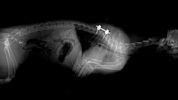
A recent abstract (ACVIM Forum 2005, Baltimore, Abstract #223, S.A. Kube et al.: Necrotizing Meningoencephalitis in Chihuahua Dogs), describes five Chihuahua dogs between ages 1 and 10 with severe acute to chronic necrotizing non-suppurative meningoencephalitis some with cystic cavitation. The dogs presented with altered mentation, seizures and blindness. The cerebrospinal fluid analysis supported a non-suppurative inflammatory process (moderate elevation of protein and mononuclear pleocytosis) and the MRI studies showed multifocal loss of demarcation between the white and the gray matter with cortical hyperintensity on T2 weighted images.
Table 1
Advertisement
All of the dogs mentioned in this abstract were euthanized (two dogs were treated prior to euthanasia using glucocorticoids and cytosine arabinoside without response). Asymmetrical multifocal necrotic areas were found primary in the gray and the white matter of the cerebral hemispheres and in the medulla oblongata in two cases.
In all of these necrotizing encephalitis cases, no etiologic agent has been identified. The search for infectious agents has been unrewarding. The disease has some similarities to granulomatous meningoencephalitis (GME) because both diseases exhibit perivascular and parenchymal mononuclear infiltrative inflammatory reaction. However, there are differences. In GME, there is an inflammatory cell accumulation (and/or proliferation) that predominates in the white matter in an angiocentric fashion while necrotizing meningoencephalitis is less angiocentric, less proliferative, and notably necrotic, involving both the white and the gray matter.
The definitive diagnosis can be made by histopathology via brain biopsy or necropsy. Currently there is no known specific treatment for necrotizing encephalitis of small breed dogs. Drugs such as corticosteroids, cytosine arabinoside and anticonvulsants (if seizures occur) can be used as empirical treatment. The prognosis is poor, and dogs are typically euthanized after a few weeks or months following the presumptive diagnosis.
Table 1 summarizes the non-infectious inflammatory diseases where necrotizing encephalitis falls as a differential diagnosis.
The small animal clinician must now consider necrotizing encephalitis when presented with a Chihuahua showing signs of cerebral dysfunction.
Dr. Nanai is a resident of the European College of Veterinary Neurology/Neurosurgery at the Animal Emergency and Referral Center in Fort Pierce, Fla.
Dr. Lyman is a graduate of The Ohio State University College of Veterinary Medicine. He completed a formal internship at the Animal Medical Center in New York City. Lyman is a co-author of chapters in the 2000 editions of Kirk's Current Veterinary Therapy XIII and Quick Reference to Veterinary Medicine.
Newsletter
From exam room tips to practice management insights, get trusted veterinary news delivered straight to your inbox—subscribe to dvm360.
Advertisement
Related Content
Advertisement


Advertisement
Advertisement
Trending on dvm360
1
Wrap up: Dog food recalled following plastic contamination complaints, and other news
2
UC Davis veterinary school adopts use of AI scribe platform
3
A new technology brings 3D-printed compounded medications to the veterinary space
4
New programs to launch at 5 top veterinary schools
5
