
|Articles|September 1, 2006
Anorectal disease: Tumors best treated by complete surgical excision
Author(s)Johnny D. Hoskins, DVM, PhD, DACVIM
Severe cases generally require both medical and surgical management.
Advertisement
Q. Please review some anorectal diseases that are seen in clinical practice today.
A. Dr. Robert J. Washabau at the 2005 American College of Veterinary Internal Medicine Forum in Baltimore gave a lecture on The Anorectum: Disease Pathogenesis, Diagnosis and Therapy. Some relevant points in this lecture are provided below.
Perineal hernia
Perineal hernia in dogs is characterized by disruption of the muscles of the pelvic diaphragm and herniation of the rectum and other pelvic organs (e.g., urinary bladder, prostate gland) into the ischiorectal fossa. Four types of perineal hernia occur, but the most-common form is the caudoventral perineal hernia, a hernia that occurs between the levator ani, external anal sphincter and internal obturator muscles.
Perineal hernia is seen almost exclusively in intact male dogs with average age of about 8 years; it occurs in female dogs on rare occasions. Perineal hernia is a less common in cats. The pathogenesis of perineal hernia appears to differ between the two species. It is associated with neurogenic atrophy of the levator ani in the dog, whereas it is often a secondary lesion associated with perineal urethrostomy or idiopathic megacolon in the cat.
Advertisement
Dogs with perineal hernia are presented with a reducible perineal swelling, tenesmus, dyschezia, constipation and/or obstipation. The perineal swelling is usually ventrolateral to the anus on one side, but severely affected dogs may have bilateral ventral swellings. Some dogs have signs of acute urethral obstruction because of retroflexion of the urinary bladder into the perineal hernial space. Tenesmus and constipation are reported often in affected cats; perineal swellings are an infrequent finding. Physical examination findings are usually straightforward. A hernial sac is palpated externally in some cases. Rectal examination reveals a defect in the pelvic diaphragm, confirming the diagnosis. Perineal hernia contents in the dog include retroperitoneal fat, serous fluid, rectum, prostate gland, urinary bladder and small intestine. Rectal wall abnormalities also may be present (e.g., rectal deviation, sacculation or diverticulum). Herniation of peritoneal contents appears to be relatively rare in cats.
Medical therapy may be attempted in mild cases of perineal hernia. About 20 percent of dogs can be maintained free of signs by the use of fecal softeners and occasional enemas. However, the owner should be advised that signs may become refractory to medical therapy, and urinary bladder retroflexion can occur at any time. Herniorrhaphy is indicated for most cases of perineal hernia. In the standard herniorrhaphy technique, the external anal sphincter is approximated to the coccygeus dorsally and laterally, the internal obturator muscle ventrally, and the sacrotuberous ligament laterally. The technical deficiencies of this method are the deformity of the external anal sphincter and the inability to obliterate large ventral hernias. The internal obturator transposition technique overcomes some of these deficiencies by elevating the obturator muscle from the ischium and suturing it to the external anal sphincter medially and to the sacrotuberous ligament laterally. The prognosis for successful repair of long-standing perineal hernias is still guarded. These dogs may suffer from rectal wall abnormalities, severe atrophy of the pelvic diaphragmatic musculature and large ventral hernias that are not easily repaired by the standard technique.
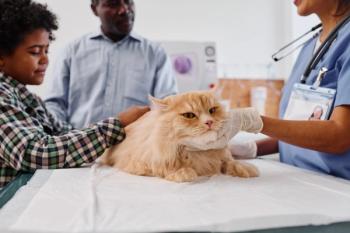
Rectal tumors
Primary tumors of the rectum are uncommon in dogs and cats. However, tumors of the colon and rectum represent 36-60 percent of all dogs and 10-15 percent of all cats alimentary tract neoplasms. The most common tumors of the rectum are benign adenomatous polyps. Polyps of the rectal mucosa are usually focal, pedunculated or sessile tumors that do not metastasize. Occasionally, these rectal polyps invade the lamina propria and submucosa and, although they appear histologically benign, are referred to as carcinoma in situ. These tumors may have a greater propensity for metastasis. Colorectal adenocarcinomas tend to spread beyond the rectal wall to regional lymph nodes, liver and lungs. Polyps and adenocarcinoma may have entirely different pathogeneses in dogs and cats. Other tumors that have been reported in the rectum include leiomyoma and leiomyosarcoma, lymphosarcoma, plasmacytoma and fibrosarcoma. Colonic and rectal lymphosarcomas are the most common large-bowel tumor of the cat. Most of these tumors occur in the ileocecal sphincter of the cat. Colorectal lymphosarcoma is less common in the dog, but most of these occur in the rectum.
Tumors of the rectum are likely to have a multitude of inciting causes. Rectal tumors are best treated by complete surgical excision. Good to excellent results are generally obtained with resections of rectal polyps or carcinoma in situ. Recurrences occur in only a few cases. Radical full-thickness resection of adenocarcinomas has been recommended, but dogs undergoing cryosurgery and local excision may have a better outcome. Wound dehiscence, infection, rectal stricture and fecal incontinence are significant complications associated with radical excision. The prognoses for rectal polyps, carcinoma in situ, leiomyomas and fibromas are generally favorable. Adenocarcinomas, lymphosarcomas and plasmacytomas tend to recur. Dogs with annular colorectal adenocarcinomas have a particularly poor prognosis with average survival time of only 1.6 months.
Perianal fistula
Perianal fistula, or anal furunculosis, is a chronic debilitating condition of dogs characterized by single or multiple ulcerated sinuses that can involve up to 360 degrees of the perianal tissue. True fistulous tracts from the anorectal canal to the perianal skin are uncommon in the dog, yet the term perianal fistula has persisted. Dogs with perianal fistula are typically presented with tenesmus, dyschezia, fecal incontinence, licking of the anal area, anal bleeding, constipation and/or a malodorous anorectal discharge. Anorexia and weight loss also can occur in some dogs with severe ulceration or infection. Large-breed dogs are most commonly affected, with the highest incidence in German Shepherds and Irish Setters. Physical examination of the anorectum may require sedation or anesthesia in some dogs because of extreme pain. Examination usually reveals single or multiple areas of ulceration, fistulous tracts and a purulent exudate with frank hemorrhage. Anorectal palpation may reveal multiple rectocutaneous fistulas and anal stenosis.
Ruptured anal sac abscess and perianal adenocarcinoma are the only important differential diagnoses. Anal sac rupture is usually seen as a unilaterally draining tract located ventrolateral to the anus. The cellulitis and fistulation associated with anal-sac rupture is usually less extensive than with perianal fistula. The anal sacs may also be involved in animals affected with perianal fistula, rendering definitive diagnosis difficult. Perianal adenocarcinoma usually has a proliferative raised appearance, although ulceration is common, and they may grossly resemble perianal fistula.
Severe cases generally require both medical and surgical management. The principles of medical management include removal of perineal hair, cleansing and debridement; antibacterial therapy; immunosuppressive therapy; and dietary therapy. Novel antigen diets may affect clinical improvements in some cases, but complete resolution generally requires the concurrent use of immunotherapy. Immunosuppression with azathioprine, cyclosporine or tropical tacrolimus has proved useful in inducing remission in severely affected cases. Thereafter, periodic use of the immunosuppressive drugs may be required for control.
What's your question? Send your pediatric/geriatric related questions to: Pediatric/Geriatric Protocol, DVM Newsmagazine, 7500 Old Oak Blvd., Cleveland, OH 44130. Your questions will be answered by Dr. Hoskins in upcoming columns.
Dr. Hoskins is owner of DocuTech Services. He is a diplomate of the American College of Veterinary Internal Medicine with specialities in small animal pediatrics. He can be reached at (225) 955-3252, fax: (214) 242-2200, or e-mail:
Advertisement
Related Content
Advertisement


Latest CME


Advertisement
Advertisement
Trending on dvm360
1
Clinic center: New $15 million animal shelter proposed for Ellis County, Texas, following SPCA closure; Chewy Vet Care opens first North Florida practice in Jacksonville; and more
2
Advanced Modalities in Wound Management: Evidence-Based Strategies Through the 4 Stages of Wound Repair
3
Q&A: SGLT2 inhibitors in feline diabetes and what to know about blood ketones, DKA, diet, and monitoring
4
The next casualty of the overdose crisis could be wildlife conservation
5