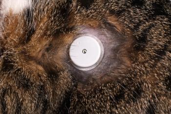
Adrenal disease in cats (Proceedings)
Although only recently discovered, feline adrenal disorders are becoming increasingly more recognized. Feline adrenal disorders include diseases such as hyperadrenocorticism (Cushing's syndrome) and hyperaldosteronism (Conn's syndrome).
Although only recently discovered, feline adrenal disorders are becoming increasingly more recognized. Feline adrenal disorders include diseases such as hyperadrenocorticism (Cushing's syndrome) and hyperaldosteronism (Conn's syndrome). The clinical signs of feline hyperadrenocorticism, which include unregulated diabetes mellitus and severe skin atrophy, are unique to the cat. Other signs of feline hyperadrenocorticism, such as potbellied appearance, polydipsia, polyuria, and susceptibility to infections are also seen in dogs with hyperadrenocorticism. Conn's syndrome has only recently been described in the cat and is in fact more common in cats than in dogs. Characterized by severe hypokalemia, hypertension, and muscle weakness, Conn's syndrome may be misdiagnosed as renal failure. The clinician should become familiar with the clinical signs of adrenal disorders in cats and the common diagnostic tests used to diagnose and treat these syndromes.
Feline Cushing's syndrome (FCS) is a disorder of excessive cortisol secretion by the adrenal glands. FCS is most often caused by a pituitary adenoma with subsequent corticotrophic hyperplasia and excess adrenocortical cortisol secretion. Also found in cats with FCS are autonomously functioning benign adenomas or malignant adrenal carcinomas. Iatrogenic FCS due to glucocorticoid administration is rare in cats. Differential diagnoses include diabetes mellitus, insulin resistance, acromegaly, hepatopathy, renal disease, sex hormone-secreting adrenal tumors, and yperthyroidism.
Pituitary-dependent hyperadrenocorticism (HAC) is usually a disease of the middle-aged to older cat. There is a slight difference in sex distribution in feline HAC; female cats are slightly more likely to develop the disease than males. No breed predilection has been found.
Feline HAC is usually (80%) accompanied by diabetes mellitus (DM). The most common clinical signs associated with HAC in cats are insulin-resistant DM, cutaneous atrophy, polydipsia, polyuria, polyphagia, lethargy, abdominal enlargement or potbelly, panting, obesity, muscle weakness, and recurrent upper respiratory and urinary tract infections. On physical examination, the most commonly noted abnormalities include abdominal enlargement, hepatomegaly, bilaterally symmetric alopecia, cutaneous atrophy with open sores, and seborrhea. Lethargy (dullness) has been reported due to muscle weakness or the effects of a pituitary mass. Excess sex hormones, such progesterone, have also been identified in cats with FCS.
In the dog, the most common serum chemistry abnormality observed in association with HAC is an increased serum alkaline phosphatase activity (ALP), which is high in most dogs. However, in the cat serum ALP is not elevated because of hypercortisolemia but rather is a result of poorly regulated concomitant DM. This occurs because cats lack the glucocorticoid-induced isoenzyme for ALP. High serum alanine transferase activity (ALT), hypercholesterolemia, hyperglycemia, and low blood urea nitrogen (BUN) are also common findings. The hemogram may reveal a mild erythrocytosis as well as a classic "stress leukogram" (i.e., eosinopenia, lymphopenia, and mature leukocytosis).
Although in dogs with HAC, the urine specific gravity is usually less than 1.015, cats often show concentrated urine specific gravity (>1.030) despite profound polydipsia and polyuria resulting from the concurrent DM. Finally, many cats with HAC have evidence of urinary tract infection without pyuria (positive culture), bacteriuria, and proteinuria resulting from glomerulosclerosis.
Screening tests
The urine cortisol: creatinine ratio (UCCR) is a simple and valuable screening test for HAC in cats. To perform this test, the owner is instructed to collect morning urine samples from an empty litter box at the same time of day on two to three consecutive days. Special precautions are needed for the urine collection itself (no contamination with litter) and the urine samples should be kept refrigerated. This home-collection protocol avoids the "stress" of a visit to the veterinary clinic.
The low-dose dexamethasone suppression test is considered by many to be the test of choice for the diagnosis of HAC in cats. It requires 10 times the dose used in dogs or 0.1 mg/kg IV. Plasma is obtained for cortisol concentrations before, 4 hours after, and 8 hours after dexamethasone administration. If the low dose of dexamethasone (0.1 mg/kg) fails to adequately suppress circulating cortisol concentrations in a cat with compatible clinical signs, this is consistent with a diagnosis of HAC. Normal cats and cats with nonadrenal illness will show adequate suppression of serum or plasma cortisol at 4 and 8 hours post dexamethasone administration. However, in contrast to dogs with pituitary dependent hyperadrenocorticism (PDH), many cats with PDH will not suppress at 4 hours and a few cats will suppress at 8 hours after dexamethasone administration.
The corticotropin ACTH stimulation test, mainly a test of adrenal reserve, requires little time, is easy to interpret, and can be used to document iatrogenic HAC. Only 50 to 60% of cats with HAC have an exaggerated response to ACTH administration. The preferred method for ACTH stimulation testing in cats is to determine serum cortisol concentrations 30 minutes before and 1 hour after the intravenous or intramuscular injection of cosyntropin.
Differentiation tests
The high-dose dexamethasone suppression test (HDDST) is performed by administering a high dose of dexamethasone (1 mg/kg IV) in a protocol identical to the low-dose dexamethasone suppression test (LDDST). An at-home version using multiple UCCRs and oral dexamethasone is easier to perform and interpret than the in-hospital protocol. The at home dexamethasone suppression protocol is to have owners collect morning urine sample on days one, two and three. Give oral doses of dexamethasone (LDDS = 0.1 mg/kg or HDDS = 1 mg/kg) and six hour intervals for two days. Submit all three urine samples for UCCR ratio. Days one and two are basal values. Day three suppression is not seen in 50% of cats with ATH but may be seen with PDH.
Endogenous ACTH concentrations are normal to high in cats with pituitary-dependent HAC. Whereas ACTH concentrations are usually low or undetectable in cats with adrenal tumors or with iatrogenic HAC. Samples for accurate endogenous ACTH concentration determination must be collected in EDTA tubes and centrifuged immediately; the plasma is then placed into plastic or polypropylene tubes (ACTH will stick to glass) and immediately frozen until the assay is performed.
Imaging
As with the dog, ultrasonography can be used to differentiate between PDH from adrenal tumors in the cat. Symmetric adrenal glands of normal or enlarged size are suggestive of PDH, whereas unilateral enlargement supports ATH. With abdominal ultrasonography, small or noncalcified unilateral adrenal tumors can generally be readily detected, and bilateral adrenal enlargement can be visualized in cats with pituitary-dependent HAC. Ultrasonography may, in addition, detect the presence of liver metastasis or invasion of the vena cava from an adrenal carcinoma. The contralateral adrenal gland would be expected to be small in cats with unilateral cortisol-secreting tumor due to the fact that pituitary ACTH secretion has been chronically suppressed. Computed tomography (CT) and magnetic resonance imaging (MRI) are reliable methods to image either the adrenals or the pituitary glands. As with dogs, bilateral adrenal enlargement can be readily differentiated from a unilateral adrenal tumor in most cats. CT or MRI is most helpful in diagnosis of pituitary tumors; however, MRI provides superior soft-tissue contrast as compared with CT and also is more accurate for visualization of smaller pituitary tumors.
Treatment
Medical therapy has been successful in some cats with HAC; however, the majority of cats do not respond to mitotane. Trilostane is an orally administered competitive inhibitor of 3-betahydroxysteroid dehydrogenase, the enzyme that mediates the conversion of pregnenolone to progesterone and, hence, its end-products (cortisol, aldosterone, and androstenedione) in the adrenals. Studies in cats with HAC have shown that trilostane is an effective steroid inhibitor that is associated with minimal side effects. Trilostane is administered at a dosage of 30 to 60 mg per cat per day. Trilostane therapy is monitored with weekly ACTH stimulation tests.
Surgical therapy
Surgical treatment of feline HAC consists of unilateral or, more commonly, bilateral adrenalectomy. Management of the cat during the preoperative, operative and postoperative period is essential for a good outcome. Before adrenalectomy, the cat should be regulated on trilostane until the skin lesions of HAC have resolved and DN, if present, is reasonably well controlled (no ketones). With bilateral adrenalectomy, glucocorticoid and mineralocorticoid supplementation should be initiated immediately before adrenalectomy and monthly thereafter for the rest of the cat's life. Complications following adrenalectomy include dehiscence, poor wound healing, Addisonian crises, and enlargement of the pituitary tumor, which may result in blindness or seizures (Nelson's syndrome). Response to bilateral adrenalectomy is usually good, with most cats having a resolution of clinical signs in 2 to 4 months. In approximately 50% of cases, DM resolves completely and in the other 50% insulin requirements are decreased dramatically. Transsphenoidal hypophysectomy for the treatment of feline PDH is a viable alternative to adrenalectomy however this procedure is being performed in only a few clinics in the US.
Radiation therapy
Because approximately 85% of cats with HAC have PDH, radiation therapy is another treatment option for many patients; however, radiation therapy is expensive and time-consuming. Radiation therapy is an effective method of treatment in cats associated with low morbidity, but signs of PDH may take several months to subside in treated animals. The major advantage of pituitary irradiation is that the primary disorder, a pituitary tumor, has been addressed. Cats with FCS that undergo bilateral adrenalectomy followed by pituitary irradiation have the best prognosis and many will live a normal life (with resolution of the DM) following these procedures.
Feline hyperaldosteronism
Feline hyperaldosteronism may be caused by a unilateral aldosterone-secreting adrenal tumor or bilateral adrenal hyperplasia. Tumors of the adrenal are usually benign; however, reports of an adrenocortical carcinoma secreting aldosterone have been described. Over secretion of aldosterone results in the classic electrolyte changes of hypokalemia, hypernatremia, and metabolic alkalosis (opposite of Addison's disease). However, primary hyperaldosteronism and secondary hyperaldosteronism caused by renal disease may be difficult to differentiate.
Hyperaldosteronism is associated with clinical signs resulting from systemic hypertension caused by expansion of blood volume or by polymyopathy resulting from hypokalemia. There is no breed predilection; however, reported cases tend to occur in older cats. In a report of 13 cases of primary hyperaldosteronism in cats, the most common clinical sign was hypokalemic polymyopathy, presenting as ventroflexion of the neck, paresis and hind limb weakness in cats. Less commonly, hypertension, fundic changes, blindness, polydipsia and polyuria, and polyphagia were observed.
The most common laboratory findings were moderate to severe hypokalemia in all cats; elevations in serum creatine kinase were observed in the majority. Only one cat exhibited hypernatremia and no cats showed metabolic alkalosis (a characteristic of hyperaldosteronism in human beings). Also surprising was the low incidence of azotemia in these cats with only two cases showing elevations of both serum creatinine and BUN. Urine-specific gravity was normal in most of the cats; however, two cats did show isosthenuria. Elevated plasma aldosterone concentrations were measured in all cases.
Plasma renin activity (PRA) was not measured in any of the cases. Abdominal ultrasonography revealed unilateral adrenal enlargement (1-3.5 cm) with an adrenal mass in almost all cases, all of which were biopsied and diagnosed as adrenal adenomas (seven cats) or carcinomas (six cats); two cats had bilateral adrenal enlargement with adenomas on postmortem examination.
Treatment of cats with primary hyperaldosteronism resulting from a unilateral adrenal tumor consists of potassium supplementation, an aldosterone blocker such as spironolactone, and amlodipine. None of the reported cases showed a normalization of serum potassium with supplementation; however, all cats showed resolution of the clinical signs of hypokalemia. Most hypertensive cats became normotensive on calcium channel therapy. Three cats were treated medically and eventually euthanatized due to chronic progressive renal failure. Surgical removal of the adrenal mass has been considered the treatment of choice in most cases.
In cats with primary hyperaldosteronism caused by benign bilateral adrenal hyperplasia; hypertension, blindness, and renal failure are more common than signs of hypokalemia (i.e., muscle weakness, cervical ventroflexion, and paresis). In a study of 11 cats with primary hyperaldosteronism, the cats were more likely to be older and have higher systolic blood pressure than the cats with adrenal tumors. Many of the affected cats exhibited ocular signs of hypertension such as retinal hemorrhage, hyphema, retinal detachments, and blindness; in contrast, only two cats with adrenal tumors exhibited blindness as a clinical sign.
Classic laboratory abnormalities observed in primary aldosteronism such as hypokalemia, elevated CK, and metabolic alkalosis are less commonly observed in cats with bilateral adrenal hyperplasia. Hypernatremia was not observed in cats with bilateral adrenal hyperplasia. Azotemia was observed in most of the cats. In the case of bilateral adrenal hyperplasia, diagnosis was achieved by documentation of increased plasma aldosterone, low to undetectable PRA, and/or increased plasma aldosterone concentration (PAC) to PRA ratios. In contrast to the previous study of adrenal tumors producing aldosterone, only four cats with bilateral adrenal hyperplasia had elevated PAC. PRA was measured in all cats in this study and was found to be abnormally low in most of cats. However, all cats showed a high PAC: PRA ratio.
Because idiopathic adrenal hyperplasia resulting in hyperaldosteronism is a bilateral disease, medical treatment is the only option available. Treatment of cats with primary hyperaldosteronism resulting from a bilateral adrenal hyperplasia consists of potassium supplementation, an aldosterone blocker such as spironlolactone, and antihypertensive therapy such as amlodipine or beta-blockers (atenelol). Most of the cats with bilateral adrenal hyperplasia eventually succumb to progressive renal insufficiency.
References available on request
Newsletter
From exam room tips to practice management insights, get trusted veterinary news delivered straight to your inbox—subscribe to dvm360.


