

|Articles|July 21, 2021
- dvm360 August 2021
- Volume 52
The digital cytology scanner: A revolutionary innovation
Author(s)Steve Dale, CABC
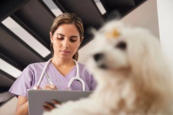
With the release of Antech’s and IDEXX’s in-clinic digital cytology scanners, veterinary professionals can deliver fast results to clients and connect with pathologists like never before.
Advertisement
Veterinary medicine has long eclipsed human medicine in terms of the speed of diagnostic results and return calls to clients. Most recently, in-clinic digital cytology scanners from Antech and IDEXX have been further speeding the process and adding efficiency to the industry by yielding fast results, connecting veterinarians to pathologists, and improving client adherence and satisfaction. Learn more about the benefits of these scanners and their technology as outlined below.
The accuracy of digital cytology scanners
Excellent images allow pathologists to interpret with ease. No significant differences have been noted between digital and glass slides regarding the number of cases correctly diagnosed or the sensitivity, specificity, or diagnostic accuracy, irrespective of the observer’s expertise. Digital slides produced by the whole-slide imaging scanner are adequate to diagnose cytological samples and are similar among clinical pathologists with differing levels of expertise.1 Additionally, when it comes to comparison of human to veterinary implementation of digital pathology, guidelines for validating scanners are certainly more than comparable on the veterinary side.2
Faster results
Whole-slide interpretations from diagnostic labs Antech and IDEXX have a rapid turnaround time of 2 hours—24/7, 365 days a year. Although IDEXX launched its in-clinic cytology reader at the Veterinary Meeting & Expo in 2020, Antech’s was launched earlier this year.
Sarah Johnson, DVM, MS, DACVP, assistant chief medical officer of clinical pathology at Antech, said, “We’ve been reading digital hematology for years and we have now transitioned to digital cytology, both at the reference lab level and with our in-hospital digital cytology scanners. There were some absolute necessities before we could feel confident with our units; they are user friendly, quick to scan, able to easily pick up on intricate details with image quality similar to a microscope, and there are no parts that can easily break, such as a slide loading cartridge.”
Penny Guyton, senior director of global product management for IDEXX Reference Labs, said, “Our customers have been so impressed, particularly during the pandemic when clients are waiting curbside in cars or dropping off a pet, and then by the time they run some errands the cytology is back. Veterinarians feel good about this—offering answers quickly.”
Advertisement
IDEXX and Antech are able to manage this 2-hour-or-less turnaround time even in the wee hours because a pathologist does not receive a 3 AM wake-up call from a criticalist in New York City. Instead, a pathologist in Australia may read the results in real time.
Easier communication
Speed is not the only benefit of the digital cytology scanners. Another advantage is the pathologist’s ability to solicit a second pathologist’s opinion nearly instantaneously.
Zachary Wright, DVM, DACVIM (oncology), chair of the VCA (Veterinary Centers of America Inc) Pet CancerCare Alliance, said, “The [Antech] cytology scanner has dramatically improved the efficiency in our workflow. We now often call clients with cytology results before they get home from their appointment. Also, for intraoperative cases it allows for a more accurate diagnosis in challenging cases that require the clients to make life-or-death decisions quickly. Lastly, the digital nature of the cytology allows for sharing among pathologists and I often get 3 or 4 opinions on challenging cases, which improves our confidence in the diagnosis.”
Client adherence and satisfaction
Additionally, client adherence has improved. Guyton pointed out that in some rural places it is ideal to have clients remain in the parking lot or in the clinic with the pet until the results return because their ride home takes 2 hours. With the client remaining in the vicinity when the results come back, treatment can start or at least a treatment plan can be created.
Johnson offered a real-life example. “A dog comes in for a wellness visit, and the veterinarian notes a lymph node should be aspirated. This is, of course, common. The dog didn’t seem ill, and indeed has no signs. Rather than say, ‘We’ll just watch and wait until the next visit’ (arguably not the best medicine) or ‘I’ll let you know in a couple of days,’ when results are going to come in so fast, clients are—by human nature—more likely to say, ‘Go ahead [with treatment].’ And if treatment is suggested, that treatment can start now. Certainly, this is to a pet’s benefit.”
The nearly instant results also may help clients sleep better—instead of returning home with their pet wondering and waiting for the results. And even if the news from the cytology is not great, the veterinarian can explain the next steps face to face.
Johnson said sometimes clients are ready to euthanize a beloved pet. “Here’s something I’ve seen: the in-clinic cytology scanners saved lives. A dog is vomiting and has enlarged lymph nodes all over. I’m thinking lymphoma, but the cytology says otherwise, and it turns out to be salmon poisoning disease3 (one example of a medical condition often mistaken for lymphoma [but not nearly as costly as treating lymphoma]).” Therefore, clients are more likely to go forward with treatment rather than euthanizing their pet due to factors such as cost, age, the progression of the disease, the scary cancer world, etc.
The future of cytology
Guyton said, “The in-clinic scanners are to everyone’s benefit; the veterinary clinic feedback we’ve received, so far, is 100% positive, that’s 100%. But also, clients, of course, want instant answers and they get it. Most important, the pets benefit. This is a path on the way to AI [artificial intelligence] for sure. This [technology] is ripe for AI, and exciting to see what’s next.”
References
- Bonsembiante F, Bonfanti U, Cian F, Cavicchioli L, Zattoni B, Gelain ME. Diagnostic validation of a whole-slide imaging scanner in cytological samples: diagnostic accuracy and comparison with light microscopy. Vet Pathol. 2019;56(3):429-434. doi:10.1177/0300985818825128
- Pantanowitz L, Sinard JH, Henricks WH, et al; College of American Pathologists Pathology and Laboratory Quality Center. Validating whole slide imaging for diagnostic purposes in pathology: Guideline from the College of American Pathologists Pathology and Laboratory Quality Center. Arch Pathol Lab Med. 2013;137(12):1710-1722. doi:
10.5858/arpa.2013-0093-CP - Booth AJ, Stogdale L, Grigor JA. Salmon poisoning disease in dogs on southern Vancouver Island. Can Vet J. 1984;25(1):2-6.
https://pubmed.ncbi.nlm.nih.gov/17422349
Articles in this issue
over 4 years ago
Case report: canine spinal cord nephroblastomaalmost 5 years ago
A whole new world’: Navigating the pandemic era in veterinary medicinealmost 5 years ago
Extraocular myositis in a young pit bullalmost 5 years ago
Minimalist design, maximum effectalmost 5 years ago
Compliance for canine mitral valve disease therapy just got easieralmost 5 years ago
IVSA and Vetstream announce a collaborative partnershipalmost 5 years ago
Time for topicals: A spot-on guide to treating dermatologic diseasesalmost 5 years ago
Pet Releaf debuts professional product line with SentesaAdvertisement
Related Content
Advertisement


Latest CME


Advertisement
Advertisement
Trending on dvm360
1
Tornado (the dog) touches down at the emergency department
2
Q&A with an emergency veterinarian on critical care and common ER misconceptions
3
Every 30 extra minutes under anesthesia raises complication risk in brachycephalic patients, underscoring the importance of peri-anesthetic management
4
When veterinary partnerships work (and when they don’t)
5