

|Articles|November 1, 2009
Primary hyperaldosteronism (Proceedings)
Author(s)Jana Gordon, DVM, DACVIM
Mineralocorticoids are hormones produced by the adrenal glands that regulate fluid and electrolyte balance.
Advertisement
Mineralocorticoids are hormones produced by the adrenal glands that regulate fluid and electrolyte balance. There are three hormones with mineralocorticoid activity that are made in the adrenal glands: cortisol, 11-deoxycorticosterone and aldosterone. Cortisol is produced in the zona fasciculata and, to a lesser extent, in the zona reticularis. Cortisol is highly bound to corticosteroid-binding globulin (CBG) in circulation. In the kidney, colon and salivary glands cortisol is converted to the much weaker mineralocorticoid, cortisone. Because there is little free cortisol in circulation and its conversion to cortisone, cortisol is considered a weak mineralocorticoid. 11-deoxycorticosterone is another relatively weak mineralocorticoid made in the zona glomerulosa that is also highly bound to CBG with very little free hormone available to bind mineralocorticoid receptors. Aldosterone is the most potent mineralocorticoid because of its high affinity for the mineralocorticoid receptor and large proportion of free hormone present in circulation.
Physiology
Aldosterone is synthesized in the adrenal gland from cholesterol. It is the restriction of the P450aldo enzyme (aldosterone synthase) within the zona glomerulosa that limits aldosterone synthesis to this layer. The primary stimulus for production is activation of the renin-angiotensin-aldosterone system. Renin is released from the juxtaglomerular cells of the afferent arterioles in the kidney as a result of decreased renal arteriolar pressure and sympathetic stimulation at the juxtaglomerular apparatus. Decreased sodium and chloride sensed by the macula densa in the renal tubules also stimulate renin release. Renin then converts circulating angiotensinogen to angiotensin I. Angiotensin I is converted to angiotensin II by angiotensin-converting enzyme. Angiotensin II then stimulates aldosterone secretion by the adrenal gland.
Aldosterone exerts it effects by binding to mineralocorticoid receptors in the cytoplasm and nucleus of cells. In the kidneys as a response to increased aldosterone, sodium channels in the apical membrane of the convoluted tubules and collecting ducts are opened and basolateral Na/K ATPase pumps are activated and increase in number. The net result is an influx of sodium from the tubular lumen into the cells making the tubular lumen less negative. Potassium is lost into the luminal fluid in order to maintain electrical neutrality. Because of the influx of sodium and water, vascular volume increases. The increase in sodium sensed by the macula densa and the increased vascular volume sensed by stretch receptors in the juxtaglomerular apparatus shuts off renin release. Potassium is also able to directly stimulate the adrenal gland to produce aldosterone. Aldosterone's effects on acid/base are primarily through increases in H+ secretion from the intercalated cells in the collecting ducts.
Etiology
Hyperaldosteronism may be primary or secondary. Secondary hyperaldosteronism usually occurs with severe cardiac, hepatic or renal disease. Primary hyperaldosteronism occurs as a result of an adenoma, adenocarcinoma or hyperplasia affecting the zona glomerulosa of the adrenal gland resulting in increased aldosterone production. There have been two reports of cats with adrenal tumors and increases in progesterone and aldosterone.
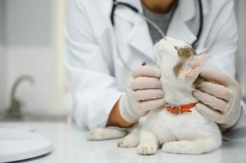
Clinical findings
Dogs and cats with primary hyperaldosteronism are middle-aged to older. Clinical signs include lethargy, weakness, anorexia, cervical ventroflexion, paresis, ataxia, forelimb stiffness, collapse and PU/PD. Hypertension is common so signs of end-organ damage might occur including hyphema, retinal detachment, hypertensive retinopathy and blindness.
Diagnosis
Common laboratory findings include hypokalemia, increased CK, increased BUN and increased creatinine. Urine is variably concentrated. A mass in the area of the adrenal(s) may occasionally be seen on radiographs but adrenal masses or enlargement are more easily detected with ultrasound or MRI. In some cats the adrenal gland(s) may be normal in size or only slightly enlarged.
Diagnosis has relied on the measurement of aldosterone and renin in plasma. As a result of increased aldosterone, renin production will decrease in normal individuals. Because there is overlap in aldosterone and renin levels in normal and affected individuals aldosterone:renin ratios and aldosterone:plasma renin activity ratios have been used to diagnose hyperaldosteronism. With hyperaldosteronism an increase in the ratio will be seen in affected individuals. An assay is not currently available to measure plasma renin activity in dogs and cats. Interestingly, plasma renin levels have been found to be normal in several cats with primary hyperaldosteronism.
Primary hyperaldosteronism is suspected in dogs and cats with the appropriate clinical signs and findings of hypertension, hypokalemia, an adrenal mass (or enlargement) and increased plasma aldosterone levels. Measurement of urine cortisol:aldosterone ratio following suppression with fludrocortisone has been suggested as a means of diagnosing primary hyperaldosteronism in the cat and may prove useful in the future.
Treatment
Surgery is the treatment of choice for animals with unilateral adrenal masses and may be elected with bilateral disease as well. If a bilateral adrenalectomy is performed supportive care for permanent glucocorticoid and mineralocorticoid deficiency must be instituted. With complete excision hypokalemia and hypertension typically resolve. With bilateral disease or in the presence of metastasis medical management may be indicated. Hypokalemia can be treated with oral potassium gluconate but potassium losses can be severe and difficult to normalize with oral supplementation. Intravenous potassium chloride is often used in hospitalized patients and is more effective than oral therapy. Spirinolactone is an aldosterone antagonist that decreases sodium and water absorption as well as potassium losses. Clinical signs of hypokalemia often improve even if potassium levels remain below normal. Most of these animals are hypertensive so treatment with calcium channel blockers and ACE inhibitors is common.
Dogs and cats that are managed medically for hyperaldosteronism may live up to 900 days. Cats treated surgically that survive the perioperative period have survival rates of over 1800 days. Some animals have existing or develop renal disease that progresses despite medical management.
References
Ash RA, Harvey AM, et al. Primary hyperaldolsteronism in the cat: a series of 13 cases. J Fel Med Surg 2005; 7:173-82.
Behrend EN, Weigand CM, et al. Corticosterone- and aldosterone- secreting adrenocortical tumor in a dog. J Am Vet Med Assoc 2005 May 15; 226(10):1662-6.
Breitschwerdt EB, Meuten DJ, et al. Idiopathic hyperaldosternism in a dog. J Am Vet Med Assoc 1985 Oct 15; 187(8):841-5.
Briscoe K, Barrs VR, Foster DF, et al.Hyperaldosteronism and hyperprogesteronism in a cat. J Felin Med Surg 2009 Sep;11(9):758-62.
Djajadiningrat-Laanen SC, Galac S, et al. Urinary aldosterone to creatinine ratio in cats before and after suppression with salt or fludrocortisones acetate. J Vet Int Med 2008; 22:1283-8.
DeClue AE, Breshears LA, Pardo ID, et al. Hyperadlosteronism and hyperprogesteronism in a cat with an adrenocortical carcinoma. 2005 May-Jun;19(3):355-8.
Flood SM, Randolph JF, et al. Primary hyperaldosteronism in two cats. J Am. Anim Hosp 1999;35:411-16.
Javadi S, Djajadiningrat – Laanen SC, et al. Primary hyperaldosteronism, a mediator of primary renal disease. Dom Anim Endocrin 2005; 28:
Machida T, Uchida E, et al. Aldosterone-, corticosterone- and cortisol-secreting adrenocortical carcinoma in a dog: case report. J Vet Med Sci 2008;70(3):317-20.
Moore LE, Biller DS, et al. Use of abdominal ultrasonography in the diagnosis of primary hyperaldosteronism in a cat. J Am Vet Med Assoc 2000 July 15; 217(2): 213-5.
Reimer SB, Pelosi A, et al. Multiple endocrine neoplasia type I in a cat. J Am Vet Med Assoc July 1 2005; 227(1): 101-4.
Rijnberk A, Kooistra HS, et al. Aldosteronoma in a dog with polyuria as the leading symptom. Dom Anim Endocrin 2001; 20: 227-40.
Advertisement
Related Content
Advertisement

Latest CME


Advertisement
Advertisement
Trending on dvm360
1
PerioVive: Regenerative Support Through Hyaluronic Acid for Every Dental Procedure
2
Weekly Vet Report: First clinical use of TMJ arthroscopy in dogs, Lyme-lepto combo vaccine approval, & more
3
New World screwworm: The flesh-eating parasite returning to the spotlight
4
Finding the right workplace culture as an LGBTQIA+ veterinary graduate
5