Nutrition: Feeding hospitalized patients for best clinical outcome
Developing a feeding plan early for your hospitalized patients can significantly increase the likelihood for recovery.
Developing a feeding plan early for your hospitalized patients can significantly increase the likelihood for recovery.
Nutrients can be supplied to the body either enterally or parenterally. Enteral (using the gastrointestinal tract) feeding provides adequate nutrition simply and cost effectively, whether done orally or by feeding tube.
Enteral feeding usually is preferred to parenteral feeding because it is less expensive, stimulates the immune system and avoids most metabolic complications. However, nutrients must be administered parenterally when the small intestine is inaccessible or not functioning adequately enough to meet the patient's nutrient requirements enterally. The two methods are not mutually exclusive. In fact, supplementing what the patient consumes voluntarily with a parenteral caloric and protein infusion is possible in most veterinary practices.
Therefore, overall patient assessment, including evaluating a patient's ability to eat and assimilate food, is the first step in developing a feeding plan because it dictates the route of administration. The route for providing nutritional support then determines which type of foods may be fed.
Oral feeding
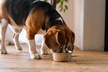
Several routes exist for enteral feeding, but the first attempt should be oral feeding unless there is a clear complicating factor, such as facial trauma. Placing a bolus of food in the proximal portion of the mouth may stimulate the swallowing reflex and, if the patient offers no resistance, is a good method as long as the patient receives enough food to meet its resting energy requirement (RER).
Simple syringe feeding of a liquid product also is a good method, if tolerated. For dogs, the syringe tip is placed outside the molar teeth and food is deposited in the cheek pouch with the head held in a normal or lowered position. For cats, the syringe tip is placed between the four canine teeth. The patient may choose to swallow the liquid or allow it to flow out of the mouth by gravity. Some patients refuse to swallow boluses of food, but force-feeding is not advisable because of the increased risk of food aspiration.
Oral feeding should be discontinued if the patient does not swallow food voluntarily. Appetite stimulants may be used to induce food consumption. However, voluntary food intake using stimulants is rarely sufficient to meet the patient's minimum caloric intake.
- OROGASTRIC TUBES require placement at each feeding but may provide a useful option for one or two days of feeding. They can be used as long as there is no nasal, pharyngeal or esophageal trauma or disease. Anesthesia or sedation is not required. Neonates tolerate multiple daily oral-tube feedings better than adults. A red rubber or polyvinyl chloride tube (8 to 24 Fr.) may be used with the tip inserted to the caudal esophagus or stomach.
An indwelling feeding tube is the method of choice if assisted feeding is necessary for more than two days. It is easier and less stressful on the patient. Nasoesophageal, pharyngostomy, esophagostomy, gastrostomy and enterostomy are potential sites. Tubes should be placed in the most proximal functioning portion of the GI tract by the least invasive method. The stomach, acting as a reservoir of a meal, should be used whenever possible.
- NASOESOPHAGEAL (NE) TUBES are generally used for three to seven days, but are occasionally used longer (weeks). Polyurethane tubes (6 to 8 Fr., 90 to 100 cm), with or without weighted tip or silicone feeding tubes (3.5 to 10 Fr., 20 to 105 cm), may be passed through the nasal cavity to the caudal esophagus or stomach.
The preferred placement of all indwelling feeding tubes originating cranial to the stomach is for the tip to be in the caudal esophagus to minimize gastric reflux and subsequent esophagitis. An 8-Fr. tube will pass through the nasal cavity of most dogs. A 5-Fr. tube is more comfortable for cats. NE feedings may be used in anorectic patients that do not have nasal, oral or pharyngeal disease or trauma.
Anesthesia or tranquilization is not necessary to place an NE tube, so this route provides a feeding option for patients considered an anesthetic risk. These tubes are most often used in the hospital, although conscientious owners can use NE tubes at home.
- ESOPHAGOSTOMY TUBE (E-TUBE) (8 to 16 Fr.) may be placed in patients with disease or trauma to the nasal or oral cavity. The tip of the tube is placed in the caudal esophagus, and the tube can be used for long-term (weeks to months) in-hospital or home feedings. For patients in which the pharynx and esophagus must be bypassed, gastrostomy tubes (G-tube) (mushroom-tipped, 16 to 22 Fr.) can be placed either intraoperatively or percutaneously using an endoscope or a G-tube introduction device.
- GASTROSTOMY TUBES are recommended also for long-term feeding (weeks to months) if needed. G-tubes are convenient and safe for in-hospital and at-home feedings. Any tube that has been placed into the esophagus or stomach allows bolus or meal-type feeding schedules because the stomach acts as a food reservoir.
- JEJUNOSTOMY TUBES (J-TUBES) (5 to 8 Fr.) are placed within the small intestine, ideally at the time of exploratory celiotomy, to bypass the proximal GI tract. J-tubes may be placed by mini-laparotomy or by threading a small feeding tube through a larger E-tube or G-tube where the tip of the smaller J-tube is in the jejunum.
A feeding tube with a tungsten-weighted tip may be threaded through the pylorus into the jejunum using an endoscope or during a surgical procedure. A common complication with J-tubes is the tip re-entering the stomach by reverse peristalsis.
Selecting a food product
Food selection depends on tube size and location within the GI tract, the availability and cost of products and the experience of the clinician. Commercial foods available for enteral use in veterinary patients can be divided into two major types: 1) liquid products and 2) blended pet foods. Nasal and jejunostomy tubes usually have a small diameter (< 8 Fr.), which requires use of liquid foods. Orogastric, E-tubes and G-tubes have large diameters (> 8 Fr.) and are suitable for liquid and blended pet foods.
Liquid foods
In general, human liquid foods cost more than veterinary liquid products and may be adequate for adult dogs, but not for long-term (> 5 days) feeding of cats, puppies and adult dogs with increased protein losses (e.g., protein-losing drains or chest tubes).
Liquid foods are of two basic types: monomeric or polymeric. Foods said to be monomeric contain nutrients in small, hydrolyzed, absorbable forms. The proteins are usually present as dipeptides or tripeptides or larger hydrolyzed protein fractions. The fat source often is an oil of mixed (medium-and long-chain) fatty acids. The carbohydrate sources are mono-, di-and trisaccharides.
These monomeric products are homogenized liquids designed for human nutrition and can be fed through any feeding tube including a J-tube. Monomeric foods are indicated in disease conditions such as inflammatory bowel disease, lymphangiectasia, refeeding parvoviral enteritis, pancreatitis and any other condition in which a patient's digestive capabilities are impaired.
Polymeric products contain mixtures of more complex nutrients. Protein is supplied in the form of large peptides (e.g., casein or whey). Carbohydrates are usually supplied as cornstarch or syrup, and fats are provided by medium-chain triglycerides (MCT) or vegetable oil.
These foods require normal digestive function and are appropriate for most veterinary clinical situations, especially when a small tube (< 8 Fr.) has been placed. Clinicare by Abbott is a polymeric product that meets the current AAFCO nutrient allowances for adult dogs and cats. This product is a homogenized liquid containing 1 kcal/ml so volume fed in 24 hrs equals RER and is usually accepted better than human liquid products containing MCT oil. This liquid food is the best option currently available in North America because of the low (230 mOsm/kg) osmolality, nutritional profile, ease of use and versatility.
Blended pet foods
This category refers to commercial products that are nutritionally complete and balanced according to AAFCO allowances for dogs and cats. Water is added typically for a consistency that flows through a feeding tube. Some products have a blended texture, high water content and very small particle size, whereas others must be blenderized with water and may have to be strained to remove particulate matter.
These products are more readily available, better tolerated and less expensive than the human liquid foods, and contain essential amino acids and micronutrients properly balanced to the caloric density of the food. Fewer medical complications (e.g., diarrhea) are likely to result.
Blended pet foods are appropriate for patients in catabolic states that are using fat and protein substrates from body stores. Blenderized canned veterinary therapeutic foods can be an asset when feeding patients with specific disease conditions requiring a specific nutrient profile (low phosphorous for example). Blenderized pet foods are more likely to plug a small feeding tube if not properly flushed after feeding; however, the patient may continue oral consumption of the same pet food, eliminating a diet change when the patient's appetite returns after the tube is removed.
The feeding schedule
Estimating a patient's approximate caloric requirement is important because feeding more of any food than is necessary may cause metabolic complications (acidosis, electrolyte shifts) and overfeeding patients through a feeding tube is possible. Diseased patients have metabolic rates and energy requirements that are less than those of comparable healthy individuals.
Requirements for all other nutrients need not be calculated when the food is "complete and balanced." When the patient consumes the proper amount of balanced food calories, all other nutrient requuirements have been met, unless known losses of particular nutrients occur (e.g., protein and electrolytes).
When it becomes certain the patient has not been eating enough food to meet at least RER for three days, then assisted feeding should be instituted and the feeding plan revised.
Calculate the RER using the current body weight because feeding for weight gain will be overfeeding a sick patient. The formula to estimate the RER of a hospitalized patient is:
RER = 70(BWkg)0.75 , or simply 15 kcal/lb dog, 20 kcal/lb cat and 25 kcal/lb of either under 5 lbs. Most hospitalized veterinary patients should be fed at their calculated RER, realizing their actual energy requirement is likely to change over the course of the disease process and recovery.
In fact, in human surgical patients, there was relatively little additional benefit to increasing intake after half of the RER requirement had been achieved. And while there are a few exceptions, initially feeding patients at RER, or slightly greater than 50 percent of RER, is a good recommendation that decreases the probability of complications. It is logistically easier to accomplish and still derives benefits of nutritional support. Feeding is preferable to starving, yet underfeeding is preferable to overfeeding.
The feeding schedule often is determined by the patient's ability to tolerate food and the logistics of feeding. Feeding an amount equal to the patient's RER during the first 24 hours of food reintroduction, if physically tolerated, is recommended. The stomach does not "shrink" during a prolonged fast, but rather the stretch receptors are more sensitive and are stimulated by a smaller volume during refeeding.
If the volume of food required to meet RER in 24 hours is not tolerated by the stomach, feeding one-third of RER and then increasing the amount by one-third every 24 hours is a more cautious approach.
Foods should be warmed to room temperature, but not higher than body temperature before feeding, and boluses must be infused slowly (over approximately 1 minute) to allow stomach expansion. Daily food dosage should be divided into several meals according to the expected stomach capacity. Gastric capacities for cats and dogs are typically 5 to 10 ml/kg body weight during initial food reintroduction after a prolonged period of no food intake.
Salivating, gulping, retching and vomiting may occur when too much food has been infused or when the infusion rate is too fast.
Some patients cannot tolerate bolus feeding to the stomach, but they benefit from a slow, continuous-rate infusion (CRI) administration (by pump or gravity flow) of a homogenized liquid food to the stomach. Ideally, homogenized liquid food should be administered through the tube using a slow, continuous drip delivered by a pump.
Foods administered through a J-tube also must be infused slowly and often in either very small quantities or by a slow gravity drip or enteral pump with an hourly rate equal to RER/24 hours because the jejunum is volume-sensitive. The patient's daily fluid requirement must be met, and additional water may be administered through the feeding tube to meet that requirement. Liquid oral medications can be administered easily through feeding tubes and then flushed. If meal feeding, each must be followed by a water-flush to clear the feeding tube of food residue. When the patient is volume sensitive, it is important to know the minimum volume required to flush the tube.
Monitoring parameters
Food intake or administration of nutritional support for hospitalized patients should be reviewed at least daily. Body weight and condition should be recorded daily; however, an animal's BCS is unlikely to change during the course of a hospital stay.
Laboratory assessments specifically for patients receiving nutritional support are generally not necessary beyond those tests already routinely performed for critically ill patients. The most common alterations that occur in laboratory parameters associated with nutrient administration are decreases in serum potassium and phosphate levels, increases in serum glucose, blood urea nitrogen and hyperlipidemia.
Even apparently stable patients might develop metabolic complications of refeeding syndrome (decreasing serum potassium and/or phosphorous). However, most patients show subjective improvement in attitude within 36 hours of refeeding when stabilized prior to refeeding.
Most parameters used to assess the nutritional status of patients will not change as a result of assisted feeding during the course of hospitalization. Laboratory parameters (e.g., albumin and total protein concentrations, RBC count and hemoglobin content) are unlikely to change in less than two weeks. The lack of measurable parameters should not be a deterrent to providing nutritional support to hospitalized patients within three days. Feeding patients early prevents protein–calorie malnutrition on the cellular level, which in turn improves outcome.
Dr. Remillard is staff nutritionist at MSPCA Angell Animal Medical Center in Boston. She received her DVM degree from Tufts University in 1987 and her diplomate certification from the American College of Veterinary Nutrition in 1991. She received specialty training as a nutrition resident at the Virginia Polytechnic Institute and did a research fellowship with the Johns Hopkins School of Medicine.
Newsletter
From exam room tips to practice management insights, get trusted veterinary news delivered straight to your inbox—subscribe to dvm360.


