New studies in veterinary internal medicine: Endocrinology
Investigating the best way to monitor dogs with hyperadrenocorticism and whether fructosamine concentrations can be used to monitor glycemic control in diabetic patients.
A wide variety of research is being done on diseases that affect small animals. Often research is initially presented as a spoken or poster abstract at specialty meetings. This research may or may not be published later. Sometimes the science isn't good enough, while at other times there isn't great interest in going on to write a full article. As such, abstracts always need to be considered less solid evidence than a published paper. Nonetheless, they often represent the true cutting edge of medicine.
At the 2014 American College of Veterinary Internal Medicine (ACVIM) Forum in Nashville, much new research was presented in the realm of veterinary endocrinology. Here is the latest on two diseases that likely affect many of your patients-hyperadrenocorticism and diabetes mellitus.
Canine Cushing's disease
GETTYIMAGES/craftvisionHyperadrenocorticism is a common endocrinopathy in older dogs. Clinical signs vary. Some may be distressing to the owner such as polyuria/polydipsia, polyphagia, panting, a pot-bellied appearance and hair loss, though these are rarely of concern medically. On the other hand, medically significant issues can occur such as proteinuria, changes to renal morphology, hypertension, hypoxemia and a thrombotic tendency. Mitotane used to be the predominant form of treatment, but trilostane has taken its place in many instances. Survival with either medication has been found to be similar. As with any therapy, it is important to understand how to monitor therapy.
Testing a lower dose of ACTH. Some of the newest research on hyperadrenocorticism was presented at the 2014 ACVIM Forum. One project of interest was to look at the dosage of synthetic ACTH we use, cosyntropin.1 After the significant price increase in cosyntropin, the cost of an ACTH stimulation test skyrocketed. Although a gel is available, the synthetic cosyntropin is preferred. The cost of ACTH can be minimized by using only 5 µg/kg rather than the previously recommended dose of an entire 250 µg vial no matter the patient's size.
In my pharmacy we reconstitute a vial and make five aliquots, which we freeze and thaw as we need, using one aliquot per 10 kg. This multicenter research trial was to determine whether a dose of 1 µg/kg was as effective as 5 µg/kg in patients suspected of having hyperadrenocorticism or in patients being treated with trilostane or mitotane. The cosyntropin was given intravenously, and blood samples were collected before and one hour after administration. Initially, the 1-µg/kg dose was given, and four hours later the 5-µg/kg dose was administered in dogs receiving mitotane or suspected of having hyperadrenocorticism. In dogs receiving trilostane, the different dosages were given on sequential days to allow timing at four to six hours after trilostane administration.
The study involved 46 dogs-26 suspected of having hyperadrenocorticism, with 12 being treated with mitotane and eight being treated with trilostane. Statistical analysis showed no significant differences in the cortisol concentrations found based on dosage. This means that a 1-µg/kg dose of cosyntropin is effective for monitoring treatment of hyperadrenocorticism as well as for diagnosing this disease. As a result, one vial of cosyntropin will be able to test many more patients than we have previously assumed. This lowers the cost of ACTH considerably, making the main cost of an ACTH stimulation test the cost of the cortisol assays.
Cortisol concentrations for monitoring therapy. When treating a patient with hyperadrenocorticism, periodic monitoring is paramount regardless of whether mitotane or trilostane is being used. Monitoring is important to determine whether dosage adjustments are needed to better control the disease or to prevent excessive adrenal gland suppression. The cost of the ACTH stimulation test has spurred interest in ways to minimize the need for this test.
A publication looked at the utility of baseline cortisol concentrations to correctly assess treatment status in dogs being treated with trilostane.2 The abstract from the article states that baseline concentrations above or equal to 1.3 μg/dl were 98% able to rule out excessive suppression, whereas values below or equal to 2.9 μg/dl correctly excluded inadequate control in 95% of dogs tested.
This sounds great and would suggest that baseline cortisol concentrations are a fantastic way to monitor these patients. When you look at the data in more detail, however, you find out that although concentrations above or equal to 1.3 μg/dl are effective when all dogs were looked at, it in fact missed 23% of the dogs that were being overly suppressed. The same applies for the 2.9 μg/dl cutoff; 17% of the poorly controlled dogs were misclassified. In addition, only 37% of the baseline cortisol concentrations were in this reference range that allowed classification. As such, the use of baseline cortisol concentrations seems questionable in most cases.
Another group of researchers performed a similar study and also concluded that the baseline cortisol concentration was not an adequate way to monitor therapy in dogs being treated for hyperadrenocorticism with trilostane.3 They did find that a cortisol concentration above 4.4 μg/dl predicted a poorly controlled dog. Their methodology was different, however, in that they performed their ACTH stimulation tests two to four hours after administration of trilostane, so this cutoff value can only be used when performing the test in this fashion. From these studies, it appears that currently we cannot avoid the ACTH stimulation test for monitoring biochemical success of therapy.
Monitoring beyond ACTH stimulation. When monitoring a patient, there is of course the adage that you don't treat the laboratory work, you treat the patient. An abstract presented at the European College of Veterinary Internal Medicine meeting in Liverpool in 2013 looked at monitoring not just from a laboratory testing point of view.4 The study encompassed 25 dogs that were being treated with trilostane. In addition to performing an ACTH stimulation test and standard laboratory tests, the researchers also had the owners fill out a questionnaire regarding the severity of clinical signs.
They found that there was no correlation between baseline or stimulated cortisol concentration and clinical signs or standard laboratory results. These results are important for us as clinicians to keep in mind when using trilostane. Dosage adjustments with trilostane should not be based solely on the results of the ACTH stimulation test. They should be based on the owner's perception of the clinical signs. If the ACTH stimulation test suggests poor control but the owner notes that clinical signs are absent, it is questionable if the trilostane dosage should be increased. Alternatively, if the ACTH stimulation test shows good control but clinical signs are still present, therapy needs to be adjusted, such as increasing the dosage or going to twice daily dosing.
Of course it is also important for us as veterinarians to monitor for those clinical changes that the owner cannot determine such as blood pressure and proteinuria. This doesn't mean we avoid doing an ACTH stimulation test. We still need this test to be sure that we are not suppressing the adrenal gland excessively.
Diabetes mellitus
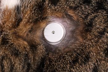
GETTYIMAGES/Uyen LeDiabetes mellitus can be a frustrating disease to manage in our patients, especially cats. It can be challenging to get reliable blood glucose curve results in the clinic, which complicates making treatment adjustments. In some cases in-home glucose monitoring can be used in the place of in-clinic monitoring, but this isn't possible in all cases. Fructosamine is a commonly used laboratory test that should indicate glycemic control over longer periods of time. In people, it gives an idea of glycemic control over the last two to three weeks. Since it is a single blood draw, it is tempting to use this parameter as a way to determine whether glycemic control is good.
Fructosamine in cats. Until recently we did not know the kinetics of fructosamine in cats. A study infused cats with dextrose to achieve long-term hyperglycemia.5 This study showed that it takes about five days for fructosamine to increase when severe hyperglycemia was induced (29 mmol/L, or 522 mg/dl) and about 20 days for maximal values to be reached. After the infusion was stopped, it took six days to return to baseline. In cats in which moderate hyperglycemia (17 mmol/L, or 310 mg/dl) was induced, the fructosamine concentration was often in the reference range, and values returned to baseline in two days.
This suggests that in cats fructosamine gives an indication of glycemic control over the last week at most and only if marked hyperglycemia is present. An earlier study had looked at ways to monitor clinical glycemic control based on various laboratory and clinical test results.6 This study found fructosamine to only be somewhat indicative of glycemic control. The study found that urine glucose, water intake and mean blood glucose determined from a 24-hour blood glucose curve were the best predictors of glycemic status.
Fructosamine in dogs. In dogs there is little published on the utility of fructosamine to determine whether glycemic control is adequate in dogs. One study showed that clinical signs and fructosamine did not correlate well, mainly because of significant overlap in fructosamine concentrations between the well- and poorly regulated diabetics.7 Research presented at the 2014 ACVIM Forum looked at diabetic dogs (some at initial diagnosis, some receiving insulin treatment), dogs with diabetic ketoacidosis and healthy dogs.8 The treated dogs were categorized as compensated or noncompensated based on clinical findings and owners' assessments of their pets.
Fructosamine was higher in all diabetic categories compared with the healthy control dogs. The dogs were categorized based on the fructosamine concentration:
• 8.3% were in the normal range (300 to 350 mg/dl)
• 11.9% had excellent glycemic control (350 to 400 mg/dl)
• 14.3% had good glycemic control (400 to 450 mg/dl)
• 14.3% had fair control (450 to 500 mg/dl)
• 51.2% had poor control (> 500 mg/dl).
Almost all treated dogs (95%) were considered to have poor control, although 70.8% were compensated based on owner perception and physical examination findings. One dog in this group did have good glycemic control based on the fructosamine concentration but was considered noncompensated clinically as it was symptomatic for diabetes at the time of examination.
It appears that in dogs fructosamine also does not accurately reflect clinical status. As such, this assay is poor at determining whether changes in insulin therapy are needed. A very low value in a patient with signs of hypoglycemia or a low blood glucose concentration on a glucose curve would be a good indicator that the insulin dosage should be reduced. Fructosamine can be assayed in conjunction with other tests including urinalysis and a blood glucose curve as another piece of the puzzle. On its own, it has little value in guiding insulin therapy.
As with hyperadrenocorticism, it is vital to get an excellent history and perform a thorough physical examination since these are much more useful in guiding therapy than the results of laboratory testing. It is good to know that veterinarians and good clinical judgment cannot be replaced by a machine just yet.
References
1. Aldridge C, Behrend E, Kemppainen R, et al. Comparison of two doses for ACTH stimulation testing in dogs suspected of or treated for hyperadrenocorticism (abst). J Vet Intern Med 2014;28:1025.
2. Cook AK, Bond KG. Evaluation of the use of baseline cortisol concentration as a monitoring tool for dogs receiving trilostane as a treatment for hyperadrenocorticism J Am Vet Med Assoc 2010;237:801-805.
3. Burkhardt WA, Boretti FS, Reusch CS, et al. Evaluation of baseline cortisol, endogenous ACTH, and cortisol/ACTH ratio to monitor trilostane treatment in dogs with pituitary-dependent hypercortisolism. J Vet Intern Med 2013;27:919-923.
4. Wehner A, Gloeckner S, Sauter-Louis C, et al. Association between ACTH stimulation test, clinical signs, and laboratory parameters in dogs with hyperadrenocorticism treated with trilostane (abst). J Vet Intern Med 2014;28:743.
5. Link KR, Rand JS. Changes in blood glucose concentration are associated with relatively rapid changes in circulating fructosamine concentrations in cats. J Feline Med Surg 2008;10:583-592.
6. Martin GJ, Rand JS. Comparisons of different measurements for monitoring diabetic cats treated with porcine insulin zinc suspension. Vet Rec 2007;161:52-58.
7. Briggs CE, Nelson RW, Feldman EC, et al. Reliability of history and physical examination findings for assessing control of glycemia in dogs with diabetes mellitus: 53 cases (1995-1998). J Am Vet Med Assoc 2000;217:48-53.
8. Claus P, Gimenes AM, Castro JR, et al. Fructosamine levels do not agree with clinical classification regarding diabetic compensation in diabetic dogs under treatment (abst). J Vet Intern Med 2014;28:1034-1035.
Anthony P. Carr, Dr. med. vet., DACVIM, is a professor of small animal clinical sciences at Western College of Veterinary Medicine, University of Saskatchewan.
Newsletter
From exam room tips to practice management insights, get trusted veterinary news delivered straight to your inbox—subscribe to dvm360.


