
|Articles|August 1, 2009
Medical management of upper airway obstruction in dogs (Proceedings)
Author(s)Lesley King, MVB, DACVECC, DACVIM
Dogs and cats with partial or complete upper airway obstruction can present with varying degrees of dyspnea.
Advertisement
Clinical signs of upper airway obstruction
Dogs and cats with partial or complete upper airway obstruction can present with varying degrees of dyspnea. Animals with complete airway obstruction may present in an agonal state and are generally quite easy to recognize! In these cases, absence of air movement is accompanied by frantic or gasping attempts to breathe with an open or gaping mouth.
Patients with varying degrees of partial airway obstruction usually have a fairly typical presentation. The hallmark clinical sign of upper airway obstruction is the presence of an audible noise during breathing. Forced movement of air through narrowed airways results in generation of loud sounds that can easily be heard without a stethoscope. Animals with laryngeal disease often make a rasping noise that is worse during inspiration, which is termed stridor. In contrast, animals with nasal, pharyngeal or soft palate disease tend to make of a snoring noise that can occur during either inspiration or exhalation, which is termed stertor. Animals with laryngeal disease often have a history of a change in sound quality of their bark.
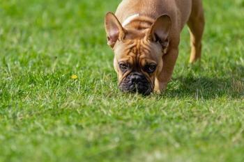
The pattern of respiration can also provide some useful information about the location of the respiratory disease. Animals with dynamic upper airway obstruction (for example laryngeal paralysis) tend to have prolonged inspiratory efforts as they try to move air into the lungs through the narrowed part of the airway. The respiratory rate may not be greatly increased above normal, but there is increased respiratory effort, sometimes associated with rhythmical opening of the mouth towards the end of each inspiration. In these animals, the expiratory phase of respiration is often completely normal as the pressure changes in the airway tend to "blow open" the larynx during exhalation. In contrast, animals with fixed upper airway obstructions, for example brachycephalic dogs or those with airway masses, tend to have trouble during both inhalation and exhalation.
The condition of animals with upper airway obstruction is often considerably worsened by stress or exercise. Even slight increases in oxygen demand can result in serious problems for some of these patients. Increased respiratory drive stimulates an increase in respiratory effort, which results in generation of greater negative pressure within the airway. Negative pressure within the airway tends to exacerbate collapse of the upper airway at the site of the obstruction, much as would happen if attempts are made to suck on a straw that is blocked at the end. The more the animal tries to inhale against the obstruction, the worse the airway obstruction becomes. A vicious cycle of progressive obstruction and dyspnea therefore ensues, that can spiral out of control within minutes.
Dyspnea can also be profoundly worsened by the presence of hyperthermia. Increased activity of the respiratory muscles, combined with stress associated with high ambient temperatures and excitement, often results in significant elevation of the body temperature of these patients. Normal thermoregulation in the dog involves evaporative cooling as water evaporates from the surface of the tongue during panting. In animals with upper airway obstruction, there may be minimal to no movement of air over the surface of the tongue, considerably restricting their ability to thermoregulate. The higher the body temperature becomes, the more they attempt to pant, resulting in generation of even greater swings in airway pressure, and further worsening of the airway obstruction. Extreme hyperthermia can have serious consequences including disseminated intravascular coagulation and shock.
Animals with upper airway obstruction are prone to the development of secondary pulmonary complications that can also exacerbate respiratory distress. The most common complication is aspiration pneumonia, particularly if the laryngeal function is abnormal and the airway is therefore unprotected. Regurgitation and vomiting are common consequences in these patients as a result of aerophagia, further exacerbating the risk of aspiration. The other common pulmonary complication is the development of non-cardiogenic pulmonary edema due to profound negative pressure within the thorax and poorly understood changes in vascular permeability.
Thus, animals with partial airway obstructions frequently present with a chronic history suggesting that airway disease has been present for a prolonged period of time. The acute presentation is often associated with a sudden and rapid decompensation to respiratory distress that is often triggered by stress, exercise, excitement, hyperthermia, or the development of pulmonary complications.
Initial stabilization
Upper airway obstruction can represent a crisis that must be managed aggressively but very carefully. The first priority is to provide immediate oxygen supplementation, using methods that will be tolerated by the patient. Dyspneic animals may not accept placement of a mask over the muzzle, and typically are not calm and stable enough for nasal cannula placement. Alternate methods that are usually reasonably well tolerated include "flow-by" oxygenation, oxygen hoods and oxygen cages. Care must be taken that the chosen method of oxygen supplementation does not result in a further increase in body temperature. Its also important to recognize that increasing the percent of inspired oxygen does not actually change the volume of air being moved past the obstruction per minute, therefore the carbon dioxide levels in the blood do not go down. Thus, although oxygenation may improve, the sensation of dyspnea in the patient is usually unchanged.
Advertisement
One of the next highest priorities is to obtain secure vascular access, if possible, without causing too much additional stress to the patient. As soon as possible, a short catheter should be placed in a peripheral vein such as the cephalic or saphenous. Obtaining vascular access allows intravenous drug administration and provides a route for crisis management should the patient decompensate or even suffer a respiratory arrest. This is of particular importance if stressful diagnostic testing such as radiography is being attempted. Placement of the catheter also is an opportunity to obtain a small blood sample from the hub of the catheter to run an emergency database consisting minimally of a PCV, TS, blood glucose and azostix. If available, venous blood gases and electrolyte determination can also provide valuable information at the same time.
Rectal temperature should be measured as soon as possible, and hyperthermia treated aggressively to minimize the additional respiratory drive generated by attempts to pant. The occurrence of heat-induced complications is directly related to the degree of temperature elevation and the duration of hyperthermia. Cooling methods can include soaking the patient with cool water, placement of ice packs, administration of cool intravenous fluids, alcohol on the pads of the feet, and placement of fans blowing cool air on the patient. The rectal temperature should be re-measured every 15 minutes, as the temperature can drop very quickly using these methods. Cooling should be discontinued once the temperature reaches 103ºF, as the temperature often continues to drop a few degrees, and hypothermia should be avoided.
Since stress and excitement also exacerbate respiratory distress, sedatives play an important role in management of these patients. Calming the animal decreases the respiratory drive and therefore partially relieves the obstruction of the airway. If possible, the animal should be placed in a quiet environment and handling kept to a minimum. Sedative drugs must be administered cautiously, however, because of the risk of complications in these unstable animals. High doses of sedatives can result in decreased blood pressure if hypovolemia is present. Similarly, excessive decreases in respiratory drive can sometimes precipitate respiratory arrest. Narcotics can sometimes cause panting which should be avoided if possible. If any sedative is to be administered, preparations should be made to deal with a respiratory or cardiac arrest, should it occur. The most effective and first line sedative in this population is usually acepromazine, administered in much lower doses than usual (start with 0.01 mg/kg IV, repeat if needed every 10 minutes up to a total dose of 0.05 mg/kg IV). Acepromazine, even if given intravenously, has a slow onset of action and will require at least 10 minutes to reach its full effect. If acepromazine alone does not provide enough sedation, the addition of diazepam (0.25-0.5 mg/kg IV) and/or butorphanol (0.2-0.5 mg/kg IV) will often have an excellent calming effect in these patients. If sedation does not result in an improvement in the patient's condition within 20-30 minutes, general anesthesia may be required (see below).
Efforts should be made to decrease inflammation within the upper airway, pharynx and larynx. Short-acting intravenous corticosteroids therefore have an important role in management of these patients. A single anti-inflammatory dose of corticosteroids rarely has serious negative effects, but caution should be exercised if aspiration pneumonia or other serious infections might be present. The author often administers a single dose of dexamethasone sodium phosphate (0.1 mg/kg IV) at presentation. Other short-acting steroids at equipotent doses may also be used.
Intravenous fluid therapy is important in animals with signs of hypovolemia. Considerable fluid losses can occur in these patients associated with hypersalivation, vomiting or regurgitation, and overheating. In addition, vasodilation as a result of hyperthermia can result in relative hypovolemia. Hypovolemic and/or distributive shock are common. The aggressiveness of fluid therapy depends on the severity of cardiovascular distress and also the presence of lung complications. If there is no lung disease, the patient can be treated with shock doses of intravenous fluids. If aspiration pneumonia or non-cardiogenic pulmonary edema are present, then fluid therapy should be considerably restricted and catecholamines such as dopamine should be used to support cardiovascular function.
Finally, in patients that are collapsed or heavily sedated, good patient positioning can significantly improve their ability to move air past an obstruction. Placement in sternal recumbency allows maximal ability to move both sides of the chest wall and optimizes ventilation:perfusion matching. Lateral and dorsal recumbency should be avoided if possible. Stretching out the head and neck decreases airway resistance in the trachea by avoiding kinking. Propping the mouth open further decreases airway resistance, as does pulling the tongue forward out of the mouth to decrease pharyngeal obstruction by the base of the tongue. This type of patient positioning can be used during the initial stabilization of the patient, and also during recovery from anesthesia.
Pre-anesthesia diagnostics and monitoring
Ultimately, investigation and hopefully resolution of upper airway obstruction usually involves anesthesia for pharyngoscopy, laryngoscopy or tracheoscopy, and possible surgery. In some animals, the severity of the airway obstruction precludes any diagnostic testing before induction of anesthesia and obtaining a secure airway. In others, some careful diagnostic testing is possible following initial stabilization. All patient manipulation should be carried out with extreme care.
Radiography is the most useful initial diagnostic test. One single lateral view of the neck and laryngeal region can sometimes provide useful information if a foreign body or mass is present, but is often quite normal in animals with functional airway obstructions. If possible, orthogonal views of the thorax should be obtained to assess for secondary pulmonary complications such as aspiration pneumonia or non-cardiogenic pulmonary edema, or related disease such as pulmonary metastasis. The animal should not be stressed to obtain the radiographs, and should never be placed in dorsal recumbency for this purpose. Sometimes, a single view of the chest with the animal in sternal recumbency is all that can be safely obtained. Other non-invasive imaging tests, including ultrasonography of the larynx, are currently being studied. Advanced imaging techniques such as computed tomography also provide very important information, especially if a mass is present, but are performed after induction of general anesthesia.
Assessment of pulmonary function using pulse oximetry and/or arterial blood gas analysis can also provide useful additional information to assess the severity of airway obstruction and lung disease, and the response to oxygen therapy. Although pulse oximetry readings can safely be obtained in most animals, arterial blood sampling can be stressful and should be avoided if it is not tolerated by the patient.
Prior to anesthesia, blood sampling for a complete blood count and serum chemistry analysis can provide useful information about concurrent illnesses, which is especially important in older or debilitated animals. Ideally, a urine sample should be obtained prior to intravenous fluid therapy to allow assessment of renal function. If significant hyperthermia has occurred, coagulation testing is appropriate to document any consumptive coagulopathy, especially if surgery is anticipated.
Many animals with upper airway obstruction present with a clinical history that includes vomiting or other gastrointestinal tract signs. In the majority of cases, resolution of the upper airway obstruction results in complete resolution of the gastrointestinal tract signs. There is usually no need to pursue additional diagnostics of the abdomen (such as radiography, ultrasound, or contrast studies) prior to addressing the upper airway disease. The exception to this is the patient that presents with airway obstruction accompanied by a distended and/or painful abdomen. Some dyspneic dogs have sufficient aerophagia that they can develop gastric distention with air that can even result in development of a GDV. Clearly, in these patients investigation and resolution of the abdominal problem becomes the first priority.
Monitoring of these patients prior to definitive resolution of their problem should be fairly intensive. Physical parameters (thoracic auscultation, observation of the respiratory rate and effort, palpation of the pulses, observation of the mucous membranes, rectal temperature) should be monitored at intervals varying from every 5 minutes to every couple of hours, depending on the condition of the patient. Continuous electrocardiography and intermittent or continuous monitoring of blood pressure provides additional important information.
Induction of anesthesia
Many animals will stabilize quickly after being managed as described above, and anesthesia and definitive management of the upper airway obstruction can be performed as an elective procedure. If the patient does not respond completely or quickly enough, however, emergency induction of anesthesia may be required.
Prior to induction of anesthesia, equipment should be gathered to ensure readiness to deal with an acute respiratory arrest, and to ensure that the animal can be intubated even in the presence of a severe airway obstruction. Emergency drugs such as epinephrine and atropine should be on hand and doses calculated in advance. A laryngoscope and a selection of endotracheal tubes should be available. A narrow diameter red rubber catheter can sometimes provide access past an obstruction for administration of oxygen in a crisis, even if the animal cannot be intubated. Finally, a tracheostomy set and a selection of tracheostomy tubes should be available in case intubation through the larynx is impossible and an emergency tracheostomy is required.
After pre-oxygenation for 5-10 minutes with a high concentration of oxygen, anesthesia is induced using short-acting injectable drugs that are minimally depressant to the cardiovascular system. Drugs that depress respiratory function are not a problem, as breathing can be controlled once the patient has been intubated. Animals with possible laryngeal paralysis are the exception, as care must be taken to avoid the use of agents that depress laryngeal function prior to laryngoscopy. Mask induction is not recommended because it is too slow in onset, resulting in a high-risk period prior to intubation. Similarly, intramuscular drug administration is not ideal for the same reason.
Because the animals have already received sedatives and are often quite debilitated, anesthetic drug doses are often lower than in healthy dogs. Choices for induction of general anesthesia include propofol titrated to effect (1-4 mg/kg IV slowly), thiopental (2-20 mg/kg IV), hydromorphone (0.1-0.2 mg/kg IV) or ketamine (4-15 mg/kg IV).
Recovery from general anesthesia should usually not be attempted unless the cause of the upper airway obstruction has been resolved, or by-passed using a tracheostomy. If the airway obstruction has not been resolved, a high-risk period occurs as the animal wakes up when he is too light to remain intubated, but still too deep to adequately dilate and protect his own airway. This is the time when most post-anesthetic respiratory and cardiac arrests occur. The same careful monitoring as pre-anesthesia should be continued during anesthetic recovery, and the patient positioned for optimal airway patency, as described above.
References available on request
Newsletter
From exam room tips to practice management insights, get trusted veterinary news delivered straight to your inbox—subscribe to dvm360.
Advertisement
Related Content
Advertisement


Advertisement
Advertisement
Trending on dvm360
1
Tarter control toothpaste receives VOHC Seal of Acceptance
2
Nonpharmacologic ways of managing pain and separation anxiety in dogs
3
Clinic center: Schwarzman Animal Medical Center completes $125 million renovation, and other updates
4
Q&A: What the 2023 ACVIM update means for leptospirosis vaccination
5