

Feature|Articles|June 8, 2026
IVDD in high-risk breeds: Early recognition, client education, and emerging preventive strategies
Fact checked by: Kristen Coppock Crossley, MA
Understanding predisposition, early clinical signs, and evolving interventions may help lower the risk of catastrophic spinal cord compression development.
Advertisement
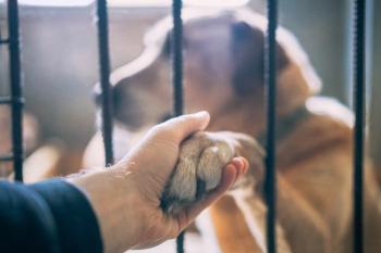
Intervertebral disc disease (IVDD) remains one of the most common neurologic emergencies encountered in canine practice and a leading cause of pain, paresis, and paralysis in chondrodystrophic breeds. Although treatment options for acute disc extrusion are well established, increasing attention has shifted toward earlier recognition of at-risk patients and strategies aimed at reducing the likelihood of future neurologic injury.
For both general practitioners and specialists, understanding breed predisposition, subtle early clinical signs, and evolving preventive approaches may provide opportunities to intervene before catastrophic spinal cord compression develops.
Understanding IVDD and chondrodystrophic breeds
IVDD refers to degeneration and displacement of intervertebral disc material, resulting in spinal cord and nerve root compression. Hansen Type I disc disease is classically associated with chondrodystrophic breeds, in which premature degeneration and mineralization of the nucleus pulposus occur at a young age.
Commonly affected breeds include the following:
- Dachshunds
- FrenchBulldogs
- Beagles
- PembrokeWelshCorgis
- Shih Tzus
- BassetHounds
- Pekingese
Recent genetic research has further clarified the association between fibroblast growth factor 4 (FGF4) retrogene expression and chondrodystrophic conformation.¹ Dogs carrying these genetic traits often experience disc degeneration significantly earlier than nonchondrodystrophic breeds.
Although thoracolumbar disc extrusion remains the most recognized presentation, cervical disc disease is also frequently encountered, particularly in smaller breed dogs.
Recognizing early clinical signs
Clinical signs of IVDD may initially be subtle and episodic. Owners often report vague behavioral changes before overt neurologic deficits become apparent. Early warning signs may include the following:
- Reluctance to jump
- Hesitation on stairs
- Intermittent kyphosis
- Cervical guarding
- Vocalization when lifted
- Reduced activity
Advertisement
- Mild ataxia
- Muscle tension or trembling
In many cases, these signs may temporarily improve with rest or anti-inflammatory medications, potentially delaying advanced imaging or specialist referral. Veterinarians should maintain heightened suspicion in predisposed breeds, particularly young-to middle-aged dogs presenting with recurrent spinal pain episodes.
One of the greatest challenges in IVDD management is that many dogs appear clinically normal until an acute extrusion occurs. Owners may attribute intermittent discomfort, reluctance to jump, or transient episodes of spinal pain to aging, overexertion, or minor injury. As a result, opportunities for earlier evaluation and risk assessment may be missed. This reality has fueled increasing interest in identifying high-risk patients before irreversible neurologic injury develops.
Clinical pearls:Identifying high-risk IVDD patients
- Do not dismiss recurrent spinal pain episodes. Repeated episodes of neck or back pain in a chondrodystrophic breed warrant further evaluation.
- Listen carefully to owner observations. Reluctance to jump, hesitation on stairs,and intermittent yelping are often early indicators of disc disease.
- French Bulldogs and Dachshunds deserve heightened vigilance.Thesebreeds commonly present with disc degeneration at a relatively young age.
- Normal neurologic examination findings do not eliminate risk. Many predisposed dogs appear neurologically normal until an acute extrusion occurs.
- Early conversations matter. Discussing IVDD risk and warning signs before a neurologic emergency develops can improve owner decision-making and referral timing.
The role of advanced imaging
Magnetic resonance imaging (MRI) remains the gold standard for diagnosing IVDD and evaluating spinal cord compression. MRI provides superior assessment of spinal cord parenchyma, disc extrusion severity, hemorrhage, edema, and concurrent lesions.
Computed tomography (CT), particularly when mineralized disc material is suspected, may also provide valuable diagnostic information and can serve as an accessible alternative in some clinical settings.
Advanced imaging not only confirms the site of compression but also frequently identifies additional degenerative discs that may place the patient at future risk. This observation has contributed to growing discussion surrounding preventive approaches in select patients.
Medical and surgical management
Treatment recommendations for IVDD depend on neurologic status, chronicity, severity of compression, and patient-specific considerations. Conservative management may include:
- Strict activity restriction
- Analgesia
- Anti-inflammatory therapy
- Rehabilitation therapy
Dogs with ambulatory pain or mild neurologic deficits may improve successfully with medical management alone. However, recurrence rates remain clinically significant, particularly in chondrodystrophic breeds.²
Surgical decompression remains the standard of care for dogs with severe spinal cord compression, progressive neurologic dysfunction, or loss of ambulation. Hemilaminectomy is the most commonly performed thoracolumbar decompressive procedure. When performed promptly, surgical intervention may result in excellent neurologic recovery in appropriately selected patients.
Preventive strategies and the emerging role of PLDA
As understanding of IVDD pathophysiology evolves, preventive strategies have received increasing attention within veterinary neurology and surgery.
Preventive recommendations may include:
- Weight management
- Maintaining lean body condition
- Activity modification during high-risk periods
- Early evaluation of recurrent spinal pain
- Rehabilitation and core strengthening programs
In select cases, prophylactic procedures may also be considered.
Percutaneous laser disc ablation (PLDA) is a minimally invasive fluoroscopically guided procedure designed to reduce intradiscal pressure by vaporizing a portion of the nucleus pulposus. The procedure is most commonly considered in chondrodystrophic dogs identified as high risk for future thoracolumbar disc extrusion.
Published studies have demonstrated a reduction in the incidence of future thoracolumbar disc extrusion following PLDA in appropriately selected chondrodystrophic dogs, although patient selection and long-term outcome assessment remain important considerations.³ The procedure is generally performed prophylactically in neurologically normal or mildly affected dogs rather than as treatment for acute severe spinal cord compression.
Because appropriate patient selection remains critical to procedural success, dogs being considered for PLDA should undergo consultation with a neurologist or surgeon experienced with the procedure to determine candidacy and discuss expected outcomes, limitations,and alternative treatment options.
Although not appropriate for every patient, PLDA represents one example of the broader shift toward preventive spinal care in veterinary medicine. Continued research and long-term outcome data will help further define the ideal role of preventive interventions in IVDD management.
Client communication and expectations
Client education remains a critical component of IVDD management. Owners of predisposed breeds often underestimate the potential severity of disc extrusion until neurologic injury occurs. Early discussions regarding breed risk, warning signs, and emergency indicators may improve response time and facilitate earlier referral.
Veterinarians should also emphasize that even successful surgical outcomes may involve prolonged rehabilitation, nursing care, and financial commitment. Establishing realistic expectations while discussing preventive health strategies can help owners make informed decisions throughout the patient’s lifetime.
Takeaway
IVDD continues to represent a major source of neurologic morbidity in dogs, particularlywithin chondrodystrophic breeds. Early recognition, timely imaging, and appropriate intervention remain essential to optimizing patient outcomes.
As veterinary medicine increasingly emphasizes preventive care, discussions surrounding spinal health and risk reduction are likely to become more common in both general and specialty practice.
By recognizing at-risk patients earlier and educating owners proactively, veterinarians may help reduce the devastating consequences associated with acute disc herniation.
Gaemia Tracy, DVM, DACVIM (Neurology),is a board-certified veterinary neurologist at Eastern Pennsylvania Veterinary Medical Center in Allentown, Pennsylvania. His clinical interests include intervertebral disc disease, minimally invasive spinal procedures, and preventive spinal health strategies in dogs.
References
- Brown EA, Dickinson PJ, Mansour T, et al. FGF4 retrogene on CFA12 is responsible for chondrodystrophy and intervertebral disc disease in dogs. Proc Natl Acad Sci USA. 2017;114(43):11476-11481. doi:10.1073/pnas.1709082114
- Levine JM, Levine GJ, Johnson SI, et al. Evaluation of the success of medical management for presumptive thoracolumbar intervertebral disk herniation in dogs.Vet Surg. 2007;36(5):482-491. doi: 10.1111/j.1532-950X.2007.00295.x
- Dugat DR, Bartels KE, Payton ME. Recurrence of disk herniation following percutaneous laser disk ablation in dogs with a history of thoracolumbar intervertebral disk herniation: 303 cases(1994-2011). J Am Vet Med Assoc. 2016;249(12):1393-1400. doi: 10.2460/javma.249.12.1393
Advertisement
Related Content
Advertisement


Latest CME


Advertisement
Advertisement
Trending on dvm360
1
ASPCA awards inaugural veterinary scholarships to students pursuing animal welfare careers
2
Creatine for veterinary professionals (Part 1): What it is, how it works, and why you should care
3
FDA launches pilot to boost US manufacturing of animal drugs
4
AAVSB tapped to develop VPA licensing examination
5