
|Articles|August 1, 2011
Introduction to pet fish medicine and surgery (Proceedings)
Author(s)Gregory A. Lewbart, VMD, MS, DACZM
The group of animals commonly referred to as "fish" is a paraphyletic group of some 30,000-plus described species (www.fishbase.org). That is, it includes all of the descendants of the common ancestor of the vertebrates (subphylum Vertebrata) with the exception of the tetrapods (subclass Tetrapoda–amphibians, reptiles, birds, mammals, totaling around 23,000 species), a fairly significant branch of the subclass Sarcopterygii alongside lungfish and coelacanths.
Advertisement
What are fish?
The group of animals commonly referred to as "fish" is a paraphyletic group of some 30,000-plus described species (
Tropical fish kept in aquaria include both freshwater and marine species. The vast majority of over 1000 species of fish kept by hobbyists are small freshwater. Marine tanks are more involved to keep and maintain, so numerically tropical marine fish comprise a much smaller portion of the pet fish population, but species diversity is comparable. Greater than 95% of freshwater fish in the pet trade are produced in aquaculture facilities, while the opposite is true for marine fish. Singapore is the world's largest exporter of tropical fish, while in the United States, Florida is the leading tropical fish aquaculture producing area, with much of the industry within an hour's drive of Tampa/St. Petersburg.
Common groups of freshwater fishes kept by hobbyists:
• Cyprinidae, the minnows, over 2000 species. Barbs, rasboras, danios, (also carp, koi and goldfish).
• Osteoglossidae, bony-tongues, 6 species. Arowanas.
• Catfishes, 15 families, over 2000 species. Including Loricaridae(including plecostomus and other "algae eaters", Callichthyidae (including Corydoras spp.), Aspredinidae (banjo catfishes), Schilbeidae (glass catfishes).
• Cichlidae, cichlids, over 900 species (over 700 from Africa). Extensive adaptive radiation of cichlids in African rift lakes has received considerable study in evolutionary biology. Also includes the South American freshwater angelfish (Pterophyllum) and discus (Symphysodon).
• Electrophoridae, electric eel or electric knifefish, one species. Requires acid water (pH 6), can breathe air.
• Mormyridae, elephant fish or elephant-nosed fish, nearly 200 species. Africa, weak electric organ, large cerebellum.
• Anablepidae, four-eyes, three species. Surface dwellers with 2 anterior chambers per eye, South America.
• Labyrinth fishes (Superorder Anabantoidei, including families Anabantidae, Helostomatidae, Belontiidae, and Osphronemidae), about 80 species. Gouramis, kissing gourami, betta. Anabantoid fishes have a suprabranchial organ called the labyrinth organ used for air breathing.
• Cobitidae, the loaches, about 110 species.
• Characidae, characins, over 1000 species. Tetras, pacu, piranha. Adipose fin.
Some representative marine fish families:
• Grammidae, basslets.
• Tetraodontidae, blowfishes
• Chaetodontidae, butterflyfishes
• Pomacanthidae, angelfishes
• Pomacentridae, damselfishes and anemonefishes
• Balistidae, filefishes
• Gobidae, gobies
• Serrandidae, groupers
• Scorpaenidae, lionfishes, scorpionfishes
• Zanclidae, Moorish idol, one species.
• Scaridae, parrotfishes
• Syngnathidae, seahorses and pipefishes
• Acanthuridae, tangs and surgeonfish
• Balistidae, triggerfish
• Ostraciidea, trunkfishes, and boxfishes
• Labridae, wrasses
Anatomy and physiology*
A knowledge of normal anatomy is a prerequisite for practicing fish medicine. Fortunately for us, the vertebrate body plan is fairly well conserved, so much of what you see if fish will be familiar, although there are some bizarre arrangements of the familiar (e.g. sea horses and flat fish), and many differences in specifics.
External anatomy.
Fins: dorsal, pectoral, pelvic, anal, caudal. Skin is thin and glandular with no stratum corneum and minimal subcutaneous layer. Contains taste buds in some species, particularly ictalurid catfish (the "swimming tongue"). Scales originate in dermis from osteogenic cells; when removed, attached epidermis is also lost. Scales are lacking in ictalurids and many eels. The head and lateral line system is a collection of porous sensory neuromasts; arrangement varies by species, most complex around the head.
Gi.
The bowel is largely as you would expect, although goldfish and carp (koi) lack a defined stomach. Pyloric cecae are present in some species. The pancreas is not distinct grossly in teleosts; pancreatic tissue may be present in liver, spleen and mesentery. The anus is located far cranial in eels.
Respiratory.
Gills are located within the opercular cavity. Gill structure: gill arches supported by bones of branchial skeleton, each with 2 rows of gill filaments (primary lamellae) bearing secondary lamellae where respiratory exchange takes place. Water flow over the secondary lamellae in counter current to capillary blood flow, resulting in incredibly efficient oxygen extraction. Gills also function in monovalent ion regulation (via specialized chloride cells) and nitrogenous waste excretion. Lungs are present in lungfish. Other air-breathing fish have other specialized respiratory organs, such as the labyrinth organ of anabantoid fish (including gouramis).
Endocrine.
The thyroid is diffuse, with follicles surrounding the ventral aorta and branchial arteries. Adrenal tissue is divided between the interrenal organ (equivalent to adrenal cortex) within the cranial kidney, and the suprarenal organ (equivalent to adrenal medulla) dorsal to the cranial kidney. Additional endocrine organs to note: ultimobranchial bodies (calcitonin), corpuscles of Stannius (electrolyte balance).
Circulatory.
The heart consists of the sinus venosus, atrium, ventricle, and bulbus arteriosus. Oxygen-poor blood is pumped from the heart through the gill capillaries for oxygenation, then to the dorsal aorta for systemic distribution, and returns via the venous system to the sinus venosus. Four distinct types of renal portal system have been described in fish.
Lymphomyeloid/hematopoiesis.
Fish have no bone marrow or lymph nodes. Hematopoiesis takes place in the cranial kidney. The spleen is the major lymphoid organ; the thymus is indistinct and paired, located under the dorsal edge of the operculum.
Urogenital.
The kidneys are located retroperitoneally along the vertebral column, and are divided functionally and to varying degrees physically into cranial (hematopoietic) and caudal (excretory) segments. The kidneys are involved in regulation of divalent ions. The paired gonads are elongated and exhibit substantial seasonal size variation.
Other.
The swim bladder is used for buoyancy control in fish that require buoyancy. Some connect with gi tract (physostomes), some do not (physoclists). Some are compartmentalized. Gas gland regulates content. Goldfish, carp, minnows, catfish, and salmonids are physostomous. Centrarchids (bass, sunfish), cichlids, and many marine species are physoclistous.
Clinical Physiology
An understanding of electrolyte and fluid balance is critical to delivery of proper supportive care. Organs involved in electrolyte balance include gills, kidneys, urinary bladder and gut. Disorders of any of these organs, plus skin wounds, can result in osmotic imbalances. Marine fish are hypo-osmotic to their environment, and therefore become dehydrated and benefit from fluid replacement therapy when organs of osmotic homeostasis are impaired. Freshwater fish are hyper-osmotic to their environment, and accumulate water and become edematous when fluid balance is impaired. Freshwater fish also may experience marked decreases in plasma osmolality in the course of a stress response (as with shipping, close confinement). In either case, osmotic stresses in freshwater fish can be ameliorated by addition of 1 - 4 ppt sea salts to the water. Elasmobranchs are a special case, maintaining plasma osmolality slightly higher than their marine environment, by tolerating high concentrations of urea. As with other heterotherms, metabolic rate of fish depends on environmental temperature. Immune response, drug metabolism, and digestion, among other things, are, therefore, temperature-dependent.
*Clinical anatomy and physiology modified from Stoskopf MK (ed). 1993. Fish Medicine. WB Saunders, Philadelphia.
The history
As with any sick animal a good history can be the clinician's best friend; be sure and obtain a complete one. The clinician will require such important information as: How long has the client owned the fish? How experienced is the client? What and how often are the fish fed? Have any new fish been introduced into the aquarium or pond recently and if so were they quarantined? Have the fish been treated with any medications? Armed with answers to these and other background questions the veterinarian or technician is ready to test the water. Developing/utilizing a medical record form tailored to the fish patient that includes the history and water quality values is recommended.
Water chemistry:
Water chemistry is the most complicated part of aquatic system management and perhaps the most important. It is necessary to have an understanding of water chemistry principles in order to successfully diagnose and correct aquarium problems.
Oxygen is the most important life-supporting component found in water. Water contains much less oxygen than air (0.004% in sea water and 21.0% in atmospheric air) and most fish must ventilate a volume of water 10 times that of the air ventilated by a terrestrial animal to obtain the same quantity of oxygen. The amount of oxygen dissolved in a given volume of water depends on four factors: temperature, atmospheric pressure, salinity and the aquatic plants in the system. In water, oxygen is either saturated, supersaturated or undersaturated. With adequate aeration from a good air pump most home aquariums are saturated and contain enough oxygen for the tank residents. Overaerating a tank can result in a condition known commonly as "gas bubble disease." In this situation, the water is supersaturated with atmospheric air and gasses enter the bloodstream and epithelial tissue of the affected fish. These emboli can kill the fish and the situation must be corrected quickly. The author has observed this problem when the tank temperature is very high (over 90° F) and the aquarist tries to compensate for the low oxygen levels by excessively aerating the aquarium. Gas bubble disease is similar to the condition known as the "bends" when nitrogen in the bloodstream of a SCUBA diver comes out of solution. Making temperature control and aeration adjustments will solve this problem. Large subcutaneous emboli may be aspirated with a syringe in some cases.
Under-saturation of aquarium water with oxygen may result in hypoxia and is fatal to fish unless corrective measures are taken. Fish experiencing this condition will commonly appear at the surface gasping for air and hyperventilating. This behavior is called "piping" by aquarists.
As temperature increases the amount of dissolved oxygen decreases. It is interesting to note that water at 4° Centigrade contains twice as much oxygen as water at 40° Centigrade. Generally speaking, cold and cool water fishes require more oxygen than warm water fishes. Two other factors to consider are pressure and salinity. As the salinity (salt concentration of the water) increases, the dissolved oxygen decreases. There is less oxygen present in seawater than in an equal volume of freshwater. As the atmospheric pressure decreases, the amount of dissolved oxygen also decreases. Thus, there is more oxygen present in a liter of Washington D.C. water than there is in a liter of Denver water with all other factors being equal. Aquatic plants and green algae also influence dissolved oxygen. During the day, photosynthesis occurs and aquatic plants give off oxygen. At night these same plants absorb oxygen, and if numerous, can deplete a body of water of available oxygen. This is rarely a problem in the home aquarium.
Tropical fish generally require between 6 and 10 parts per million dissolved oxygen to survive. Most fishes become stressed below 6 parts per million. A helpful guideline is 6-8 liters of air per hour for every 3.8 liters (1 gallon) of water in the aquarium. At this rate there will be enough oxygen for the fish, plants and nitrifying bacteria. Most established pet stores carry kits that test for dissolved oxygen if the clinician feels there may be an aeration or oxygen problem in the aquarium.
Many people speak of the importance of pH and its affect on fish health. The actual pH of the water is not nearly as important as how the pH is related to other water chemistry parameters such as ammonia. The pH is simply a measurement of dissolved hydrogen ions in the water (the symbol stands for the logarithm of the reciprocal of the hydrogen ion concentration). Most fish can survive a wide range of pH values as long as changes occur gradually. Marine fish are more sensitive to abrupt changes than freshwater fish and some species are more sensitive than others. When transferring fish from one aquatic system to another the pH values of both systems should be recorded. The normal acclimation process should include a "mixing" of the different water to allow the fish to adjust to the change in water chemistry. This is especially important if the pH gap or difference is greater than 0.5. Changes of less than 0.5 are usually not too stressful for fish but normal mixing and temperature acclimation procedures should still be employed. If the pH gap is large (e.g. 6.0-8.5) then the mixing process should be in small volume increments and for an extended period of time (an hour or more if possible). Since pH is measured on a logarithmic scale, there are 100 times as many hydrogen ions in water with a pH of 6.0 as there are in pH 7.0 water.
In unbuffered aquatic systems there tends to be a gradual lowering of the pH since hydrogen ions are given off when ammonia is oxidized to nitrate by nitrifying bacteria. This is a common occurrence in systems that are heavily loaded with fish. Most freshwater aquariums and ponds are best maintained at a pH of about 7.0 while marine systems benefit from a pH of between 8.0 and 8.5. These values are only meant to be guidelines since certain species have their own requirements. There are a number of commercially available products that will both adjust the pH and buffer the water against pH changes. Sodium bicarbonate (baking soda) can be used to increase the pH of a tank while sodium biphosphate or small quantities of hydrochloric acid may be used to decrease the pH. Anytime one decides to adjust the pH of a system with chemicals these compounds should be added carefully and gradually to avoid drastic pH changes. Experimenting first with water not containing fish, or only a few fish, is always a good idea. The buffering capacity of an aquarium will be discussed under the total alkalinity and hardness section. Aquatic systems that are not well buffered may require the addition of crushed coral, mollusk shells, or other limestone-like materials in order to increase the buffering capacity. These materials can be added to the gravel at the bottom of the aquarium or to the filtration system.
Although there is very little carbon dioxide present in atmospheric air there is a lot in natural water. After CO2 enters water, a small percentage is hydrated into carbonic acid. A portion of this carbonic acid then dissociates into carbonate and bicarbonate ions that are the primary buffers in freshwater.
Alkalinity can be defined as equivalent calcium carbonate and expresses buffering capacity. Don't confuse alkalinity with the term alkaline, a word used to describe water with a pH value greater than 7.0. While calcium carbonate is the primary buffer in water, borate (H2BO4) does account for about 5% of the total alkalinity in seawater. This buffering capacity is primarily dependent on anions (bicarbonate and carbonate) and not on cations (calcium and magnesium).
Hardness represents the total concentration of divalent cations in freshwater and is usually expressed in mg/L calcium carbonate. Calcium and magnesium are the major divalent cations associated with the carbonates (the source of alkalinity). The number of magnesium and calcium cations is frequently similar to the concentration of carbonate and bicarbonate anions. The term carbonate hardness is used to describe this condition. When alkalinity exceeds hardness some of the carbonate and bicarbonate are associated with sodium and/or potassium. When hardness exceeds alkalinity the calcium and magnesium ions are associated with anions other than carbonate and bicarbonate. In most cases freshwater hardness values are higher than alkalinity values.
Soft water (0-60 mg/L) generally has poor buffering capacity while hard water (greater than 180 mg/L) generally is a good buffer. If the hardness is high and the alkalinity is low than the previous statement will not be true. Measuring the dissolved cations in a water sample is usually an accurate but indirect way of determining buffering capacity. A good water quality test kit will be able to measure both total alkalinity and hardness.
Next to oxygen, the nitrogen compounds that are dissolved in the aquarium water are the most important factors affecting the health of fish. Most nitrogen enters a system in the form of fish nitrogenous waste. The cycling of nitrogen is primarily performed by two genera of bacteria, Nitrosomonas and Nitrobacter. Recent research has shown that other genera of bacteria contribute to nitrification in aquatic systems. These bacteria and the substrate they adhere to are called the biological filter and will be discussed later. Ammonia, nitrite and nitrate are the nitrogen compounds of importance in an aquarium system.
Ammonia found in water is generally either in the toxic unionized form (NH3) or in the non-toxic ionized form (NH4 +). The ratio between the two compounds depends on temperature, pressure, salinity and most importantly, pH. The general rule is the higher the pH, the more unionized (harmful) ammonia present. The total ammonia nitrogen (TAN) reading represents both forms of ammonia. A total ammonia measurement of 3.0 ppm would be deadly at a pH of 8.5 in freshwater but relatively harmless at a pH of 6.0 for a few days. Hobbyists and professionals commonly ask at what point should ammonia levels be considered dangerous? The best answer is that any detectable ammonia in an established system is an indicator of a filtering deficiency. Either the filter is inadequate or the biological load is too great for the filter. An elevated ammonia level combined with a low pH may keep the fish alive but the ammonia problem itself needs to be addressed or disease will develop.
Nitrite is an intermediate compound in the nitrogen cycle and is converted to nitrate by a healthy biological filter. In freshwater levels above 1.0 ppm will likely be harmful to the fish. As with terrestrial vertebrates, nitrite forms methemoglobin in the blood resulting in respiratory compromise. Affected fish will show signs of oxygen deprivation by pumping their opercula excessively and "piping" at the surface for air. Marine tropical fishes are less sensitive to elevated nitrite since the abundant chloride in seawater competes with nitrite for uptake at the gill membrane.
Nitrate is the final nitrogen compound in the nitrogen cycle and is usually not toxic to fish but persistently high levels (over 50 ppm) is probably stressful to some species. Elevated levels in an aquarium may lead to excess algal growth. Regular water changes and nitrogen monitoring will help alleviate this problem.
Introduced toxic compounds
When evaluating an aquatic system one should always find out the water source. If the water is from the tap then it should be determined whether it is city or well water. Most municipal water has been chlorinated to disinfect it for safe human consumption. While relatively harmless to humans, chlorine can be deadly to fish. The amount of chlorine in tap water may fluctuate but is usually between 0.5 and 2.0 mg/L. Chlorine can be "bubbled" out of water if the water is well aerated for several days in a container allowing for a large surface area. While effective, this method is time consuming and some people tend to lose track of time and may introduce fish to water that still contains chlorine. There are a number of commercially available compounds that instantly remove chlorine from tap water. These products usually contain sodium thiosulfate that inactivates chlorine through a chemical reaction in which sodium chloride is formed. Sodium thiosulfate is inexpensive, effective and safe. Just 7 grams of sodium thiosulfate will remove the chlorine from 1000 liters of municipal water (chlorine concentrations as high as 2.0 mg/L).
Another commonly used city water sterilizer is chloramine. This compound combines chlorine with ammonia, both of which are harmful to pet fish. The reasons behind the use of chloramine instead of straight chlorine are centered on human health concerns. Simply bubbling water or letting it stand for a week or more will not remove chloramines. Water containing chloramines must be treated with a dechlorinator like sodium thiosulfate. Your municipal water supply office can provide the interested individual with more information on how the local water has been treated.
A municipality may occasionally add copper sulfate to the drinking water in low concentrations to help control algae. Copper test kits are available in most pet stores. Levels over 0.15 ppm are dangerous to freshwater fish while levels exceeding 0.2 ppm can be harmful to marine fish. The lower the total alkalinity of the water, the more toxic copper is. Invertebrate animals such as (but not limited to) anemones, corals, crabs, shrimp, snails and sea urchins are extremely sensitive to copper. This fact must be considered if copper is being used to control a parasite problem. If high copper concentrations are consistently found in the water a special copper filter may be needed to remove this harmful compound. Copper pipes in a building's plumbing may also be a source of copper in the water.
Temperature control
Temperature is one of the easiest parameters to control in an aquatic system yet the beginning hobbyist commonly overlooks it. As a rule, freshwater tropical fishes thrive between 75 and 80 degrees Fahrenheit and marine species prefer slightly warmer temperatures (78-84 degrees Fahrenheit). Certain species of livebearers like guppies do well at room temperature and most goldfish prefer slightly cooler water (room temperature is adequate).
Water testing
Obtaining a good and durable test kit that can be purchased for between $200.00 and $300.00 is recommended. Hach Co. (
Regardless of test kit you'll want to be able to evaluate the water for dissolved oxygen, temperature, ammonia, nitrite, nitrate, pH, hardness, and total alkalinity.
Filtration
All successful recirculating systems have at least one type of filter and many have two or more. A filter is just what it says it is; it filters or removes harmful or unwanted components from the water. Several different types of filters will be discussed.
The biological filter is the best and most efficient means of removing ammonia from an aquatic system. This type of filter utilizes nitrification, a natural process that occurs constantly in soil and water. Nitrification involves the conversion of ammonia to nitrate in a two-step process. Nitrosomonas oxidizes ammonia to nitrite and Nitrobacter oxidizes nitrite to nitrate. Stable populations of these bacteria must exist in the filter for the nitrification process to perform efficiently. There are likely even more nitrifying bacterial taxa than the two genera listed. These bacteria require plenty of oxygen and ammonia as a food source. Under normal conditions it takes several weeks for the filter to develop and function adequately. When an aquarist attempts to "rush" the biological filter by loading a tank with fish before the filter is established he or she will likely be confronted with "New Tank Syndrome." This syndrome is responsible for the death of countless pet fish each year. Patience is the key when starting a new system. Begin with a few hardy fish and then gradually add more fish (properly quarantined) over time when the filter becomes established. If an aquarist just has to have a full tank in the absolute shortest period of time, then there are some commercially available biological filter starter solutions that contain nitrification bacterial cultures. Your local pet store merchant can help those interested in these products (while they probably do no harm there impact is likely negligible). The other alternative is some type of chemical filtration that physically removes the nitrogen compounds from the water. Sometimes a simple box filter containing activated carbon may be used to help remove some of the nitrogen load during the first few weeks that the system is operating. Care should be taken not to remove all of the available ammonia and nitrite or else there will be no "food" for the nitrifying bacteria.
There are several different types of biological filters. The first type is the popular undergravel filter. Most of these filters utilize a plastic grid that lies at the bottom of the tank allowing for a small water space beneath it. Several inches of gravel are then placed over the porous plate. Aerated water is pulled through the gravel bed via airlift tubes that are attached to the plastic grid. The bacteria become established on the grid/gravel and the aquarium water is literally pulled through the filter bed exposing the nitrifying bacteria to the nitrogen compounds in the water. A second type of biological filter is termed the wet/dry filter, also called an ammonia tower or trickle filter. With these filters, the bacterial bed is not submerged in water but rather is sprayed with aerated water that passes through the filter bed by means of gravity. These filters are desirable since they tend to allow for a large surface area and consequently many bacteria can colonize the filter and much ammonia can be converted to nitrate. The large municipal sewage treatment plants operate with gigantic versions of the wet/dry biological filter. Many enterprising aquarists construct their own biological filters; those with less time or mechanical aptitude can buy commercially produced wet/dry filter systems.
A mechanical filter is any type of filtration apparatus that actually strains particulates from the water. These filters usually won't remove particles smaller than 3 microns and thus will not remove ions such as ammonia and nitrite. Submerged box filters, out of tank power filters, sand filters and diatomaceous earth filters all utilize some form of mechanical filtration. Most good recirculating aquarium systems combine mechanical filtration with a biological filter.
The use of activated carbon is the most popular means of chemical filtration in the pet fish hobby. Activated carbon binds organic compounds efficiently and may act as a substitute for a biological filter. When treating a system with antibiotics or other therapeutic agents it is important to discontinue the carbon filter during treatment since the charcoal may remove the medication from the water (do not discontinue aeration).
There are two other types of filtration that have gained popularity in the past decade or so. These are the ultraviolet filter and ozone filter or ozonator. These two filters are commonly used together since ultraviolet light will inactivate ozone that can be harmful to aquatic life. These filters are not usually directly exposed to the aquarium water but rather some of the water from the aquarium is shunted to the filters so that over a period of time all of the water has passed through the filters. When water flows are properly controlled, and the filters are adequately maintained, these filters can remove organics from the water and kill both bacteria and some suspended parasites. Ozone and ultraviolet filters are very expensive and are not common in the home.
Biopsy and other sampling techniques
In many cases, and especially those dealing with a client's pet fish, the clinician may want to take some tissue samples for examination without killing the fish. Many procedures can be rapidly performed with little risk to the piscine patient. Naturally, larger fish fare better than smaller fish and the overall condition of the animal is also a factor in how it will respond to biopsy techniques.
After ruling out a water quality problem, and suspecting a parasitic or bacterial problem, one should begin with the following simple procedures: The skin scraping, fin clip and in some cases the gill snip. An anesthetic agent such as tricaine methanesulfonate (Finquel®, MS-222) can be used to restrain the fish and make these procedures easier and safer. This particular agent is purchased in a crystalline form and can be used to produce a working stock solution of 10 grams per liter of clean, dechlorinated water. By making dilutions from this stock solution the clinician can accurately formulate safe and effective anesthetic solutions. Each liter of stock solution should be buffered with about 5 grams of baking soda. The stock solution should be stored in a glass bottle away from light at room temperature. When prepared and stored in this manner, the stock solution is good for about 30 days. Final concentrations of between 100 and 150 mg/liter will usually anesthetize most fish within a matter of three to five minutes. Several studies have investigated clove oil as a fish anesthetic. Clove oil is available on an over-the-counter basis at most pharmacies. Eugenol is not completely soluble in water and should be diluted with ethanol at a ratio of 1:10 (clove oil:ethanol) to yield a working stock solution of 100 mg/ml since each ml of clove oil contains approximately 1 gram of drug. Concentrations of between 40 and 120 mg/liter are effective in freshwater and marine species and results are comparable to MS-222, except that recovery may be prolonged.
After the fish loses its ability to maintain equilibrium, it is removed from the water, the procedures are performed and the fish is placed in a "recovery" vessel containing clean water and aeration. A coverslip can be used to obtain the skin scraping sample. The coverslip should be firmly drawn across the area to be sampled, making sure that some mucus and epidermal tissue remains on the coverslip. An alternative method is to use a sterile scalpel blade for this procedure. The tissue sample that is now on the tip of the scalpel is then placed on a slide that contains several drops of clean water. After the coverslip is applied, the specimen is ready for microscopic examination. A fin biopsy can be easily obtained using a pair of fine scissors. Slide prepared with drops of water should be close by before samples are taken. Several small pieces of gill tissue can be safely cut away using small suture removing scissors after deflecting the operculum. Care must be taken to only remove a couple millimeters of primary gill lamellae. There is usually some bleeding following this procedure but it should subside quickly.
One concept to keep in mind is that many ectoparasites that affect gill tissue are usually also found on the skin and fins. Treating the parasites on the skin may also take care of the parasites on the gills. Gill damage due to environmental problems (high ammonia, bacterial gill hyperplasia) can only be evaluated after a gill biopsy has been performed.
Obtaining blood samples from tropical fish is challenging but not impossible. It is very difficult to do in fish less than three inches long (if you want the fish to survive). A sterile blood sample is a useful way to culture for a suspected bacteremia or septicemia. Fresh whole mount blood smears can be valuable in diagnosing protozoal blood parasites (e.g. trypanosomes) and in an overwhelming septicemia motile gram negative rods can frequently be observed darting across the microscopic field of view. Stained blood smears will reveal numerous nucleated erythrocytes, leukocytes and thrombocytes. Campbell and Ellis (2007) have published an excellent review of piscine hematology. Some people insert the needle along the lateral line near the tail of the fish and others take a mid-ventral approach, entering the vein from its ventral aspect. Once the needle touches the vertebral spinal body, the needle can be gently "walked" ventrally until it drops into the caudal venous sinus. Slight constant negative pressure on the plunger of the syringe will facilitate sample collection. If a blood sample is necessary from a very small fish and survival is not important in diagnosis (many other fish may be at risk and a necropsy is the best option) the tail can be removed at its base (caudal peduncle) and the small drop of blood which appears can be collected with a clean capillary tube and then placed on a slide.
Performing a fecal examination on a fish is not usually at the top of a list of diagnostic procedures even though it is a relatively easy and valuable test to perform. If the owner cannot obtain a fecal sample from the bottom of the aquarium, the fish can be placed in water containing tricaine methanesulfonate. Many fish will defecate as they relax in the anesthetic solution. If time is not a factor, the fish can be placed in a plastic bag, clean jar, or small aquarium, and a fecal sample can be collected within a matter of hours in most cases.
One final and very important area of diagnostics involves the field of microbiology. Cultures of skin and gill tissues are not especially helpful due to the ubiquitous nature of aquatic bacterial pathogens. Cultures of clinically healthy fish and clean water will commonly reveal the presence of gram negative bacteria. Clean blood samples are valuable in detecting and identifying septicemia. As with terrestrial animal medicine, every attempt must be made to procure sterile samples for microbiology. A popular culture site in fish is the kidney. The kidneys of fish run just ventral to the spinal column and are generally found the length of the body cavity. Culturing kidney tissue is a postmortem technique that is relatively easy to do. Culture samples may be processed in the clinic and antibiotic sensitivities run. Many small animal clinics are not equipped to perform microbiology testing. Fish cultures can be sent out of house since many veterinary schools and state agricultural diagnostic laboratories are familiar with fish bacteriology.
Radiology
Here at NC State radiology is part of many a pet fish diagnostic evaluation. Radiographic findings have been instrumental in diagnosing and treating a number of fish cases. Fish are relatively easy to radiograph. Most can be handled without anesthesia by simply removing the fish from a tank or bucket, placing it on a radiographic plate (protected by a plastic bag), and making the exposure. Fish will even tolerate contrast studies, CT scans, and bone scans (see below).
Ophthalmic examination
Ophthalmic problems in pet fish are commonly encountered. Diagnostic techniques such as ophthalmoscopic examination, fluoroscein staining of the cornea, and even electroretinography have been utilized to help diagnose ocular disorders of fish.
Computed tomography (CT)
This imaging modality is being utilized for fish diagnostics with more frequency and several publications describe techniques and results (see Further Reading section). As machines become "faster," imaging fish with CT becomes easier and safer since time out of the water is greatly reduced.
Magnetic resonance imaging (MRI)
Although not yet commonly used for imaging pet fish, MRI units have been installed in a number of veterinary schools and referral practices, allowing for better access and consequently more opportunities for pet fish application.
Endoscopy
This technique is gaining in popularity as more equipment and experienced operators become established. Endoscopy can be minimally invasive (exploring the oral or opercular cavities) or surgically invasive (laparoscopy). More details can be found in the Further Reading section and the University of Georgia School of Veterinary Medicine offers a short course on fish endoscopy.
Microbiology
A very important area of diagnostics involves the field of microbiology. Preparations of suspect lesions and acid-fast staining can help rule out mycobacteriosis. Cultures of skin and gill tissues may not be helpful due to the ubiquitous nature of aquatic bacterial pathogens. Cultures of clinically healthy fish and clean water will likely reveal the presence of Gram-negative bacteria. Clean blood samples are valuable in detecting and identifying septicemia. As with terrestrial animal medicine, every attempt must be made to procure sterile samples for microbiology. A popular culture site in fish is the kidney. The kidneys of most fish run just ventral to the spinal column and are generally found the length of the body cavity. Culturing kidney tissue is a postmortem technique that is relatively easy to do. Culture samples may be processed in the clinic and antibiotic sensitivities run. Many companion animal clinics are not equipped to perform microbiology testing. Fish cultures can be sent "out of house" since many veterinary schools, state agricultural diagnostic laboratories, and private laboratories are familiar with fish bacteriology.
Necropsy procedures
On some occasions, a full necropsy is the best way to arrive at an accurate diagnosis. Necropsies have several obvious advantages over biopsy procedures. Tissues can be looked at thoroughly and completely. Any and all tissues and organs are available for both gross and histopathological inspection as well as microbiological testing. If possible, the clinician should obtain a moribund fish and quickly euthanatize it for examination. An overdose of tricaine methanesulfonate (over 400 mg/liter for 15 minutes) followed by a rapid cut with a scalpel at the base of the cervical spine will quickly dispatch a fish. Once the fish is dead, the operculum can be removed to expose the gills. Samples can be taken for immediate inspection under the microscope and preserved in 10% neutrally buffered formalin for histopathology. Fish tissues can be preserved and handled like any mammalian biopsy sample. Once the abdominal organs are visible, they may be examined and removed. Small pieces of tissue may be excised, squashed on a slide under a coverslip, and examined under the microscope. This quick and easy procedure may be helpful in diagnosing a parasite problem or a condition such as hepatic lipidosis. Any suspect tissues can be preserved in formalin for histopathology. One helpful aspect of pet fish anatomy is that many animals are relatively small and entire organs or organ systems can be fixed whole in formalin.
Further reading (not inclusive)
General resources
Beleau, M.B. 1988. Evaluating Water Problems. Vet Clinics of North America, Small Anim. Pract. 18(2):293-304.
Campbell, T.W. and Ellis C. 2007. Hematology of Fish. In: Avian & Exotic Animal Hematology & Cytology. Blackwell Publishing, Ames, IA, pp. 93-111.
Evans D.H. 1998. The Physiology of Fishes (ed.) 2nd. Ed. CRC Press, Boca Raton, FL.
Gratzek, J.B, and Matthews, J.R. 1994. Aquariology: The Science of Fish Health Management. Tetra Press, 330 pp.
Hughes Hartman K., Yanong R.P.E., Harms C.A., and Lewbart G.A. 2006. The future of training for aquatic animal health veterinarians. Journal of Veterinary Medical Education, 33:389-393.
Johnson, E.J. 2006. Koi Health and Disease. Johnson Veterinary Services, 3805 Robinson Rd., Marietta, GA 30068, 204 pp.
Noga, E.J. 2000. Fish Disease Diagnosis and Treatment. Iowa State University Press, Ames, IA, 367 pp. (Note, the 2010 Second Edition has recently been published; Wiley-Blackwell, 2010, 536 pp.)
Ostrander, G.K. (Ed.). 2000. The Laboratory Fish. Academic Press, New York, NY, 2000. 678 pp.
Roberts, H.E. (ed.). 2009. Fundamentals of Ornamental Fish Health. Wiley-Blackwell, Ames, IA, 244 pp.
Saint-Erne N. 2003. Advanced Koi Care. Erne Enterprises, Glendale, AZ, 194 pp.
Spotte, S. 1992. Captive Seawater Fishes; Science and Technology. Wiley & Sons, 942 pp.
Stoskopf, M.K. 1993. Fish Medicine. Saunders Co., 882 pp. Now available via
Wildgoose W. 2001. British Small Animal Veterinary Association Manual of Ornamental Fish 2nd Edition, BSAVA, Gloucester, UK, 304 pp.
Ophthalmic examination
Bakal R.S., Hickson B.H., B.S., Gilger, B.C., Levy M.G., Flowers J.R., and Khoo L. 2005. Surgical removal of cataracts due to Diplostomum species in Gulf sturgeon (Acipenser oxyrinchus desotoi). Journal of Zoo and Wildlife Medicine, 36(3):504-508.
Jurk I. 2002. Ophthalmic disease of fish. Veterinary Clinics of North America: Exotic Animal Practice, 5(2):243-260.
Imaging
Bakal R.S., Love N.E., Lewbart G.A., and Berry, C.B. 1998. Imaging a spinal fracture in a kohaku koi (Cyprinus carpio): Techniques and case history report. Veterinary Radiology & Ultrasound, 39(4):318-321.
Eshar D., Latney L., Wyre N.R. 2009. Diagnostic contrast radiography in fish. Lab Animal, 38(10):323-324.
Govett P.D., Olby N.J., Marcellin-Little D.J, Rotstein D.S., Reynolds T.L., and G.A. Lewbart. 2004. Stabilisation of scoliosis in two koi (Cyprinus carpio). The Veterinary Record, 155(4):115-119.
Harms C.A., Bakal, RS, Khoo, LH, Spaulding, KA and GA Lewbart. 1995. Microsurgical excision of an abdominal mass in a gourami. JAVMA, 207(9):1215-1217.
Love NE, and Lewbart, G.A. 1997. Pet fish radiography: technique and case history reports. Veterinary Radiology & Ultrasound, 38(1):24-29,
Gumpenberger M. 2002. Ultrasound diagnosis—ultrasonographic detection of spinal malformation in a chub (Leuciscus cephalus L.). Veterinary Radiology & Ultrasound, 43(6):584-585.
Gumpenberger M., Hochwartner O., and Loupal G. 2004. Diagnostic imaging of a renal tumor in a red oscar (Astronotus ocellatus Cuvier, 1829). Veterinary Radiology & Ultrasound, 45(2):139-142.
Lewbart G.A., Spodnick G., Love N.E., Barlow N., Geoly F., Bakal R. 1998. Surgical removal of an undifferentiated abdominal sarcoma from a koi (Cyprinus carpio). Veterinary Record,143:556-558.
Endoscopy
Hernandez-Divers S.J., Bakal R.S., Hickson B.H., Rawlings C.A., Wilson G.H., Radlinsky M., Hernandez-Divers S.M. and Dover S.R. 2004. Endoscopic sex determination and gonadal manipulation in Gulf of Mexico sturgeon (Acipenser oxyrinchus desotoi). Journal of Zoo & Wildlife Medicine, 35: 459-470.
Murray M.J. 1998. Endoscopy in Fish. In Murray MJ, Schildger B, and Taylor M (eds), Endoscopy in birds, reptiles, amphibians, and fish. Endo-Press, Tuttlingen, Germany, pp. 59-75.
Necropsy/pathology
Ferguson H.W. 1989. Systemic Pathology of Fish. Iowa State Press, Ames, IA, 263 pp.
Plumb J.A. 1999. Health Maintenance and Principal Microbial Diseases of Cultured Fishes. Iowa State Press, Ames, Iowa, 328 pp.
Roberts R.J. 2001. Fish Pathology, Third Ed. WB Saunders, Philadelphia, PA, 472 pp.
(A version of the diagnostics/water quality section originally appeared in the 2007 AVMA Annual Conference Proceedings and have been modified and updated from: Lewbart, G.A. 1992. Basic diagnostic procedures for tropical fish. Journal of Small Exotic Animal Medicine, 1(4):182-187.)
Viral diseases
(Portions updated from Lewbart. 2005. North American Veterinary Conference Proceedings)
Several viral diseases have been thoroughly described in freshwater ornamental fishes. The most commonly observed viral disease of tropical fish is called lymphocystis disease. This disease is caused by Lymphocystivirus, an iridovirus, which infects connective tissue cells of the fish. The virus induces these cells to undergo extensive hypertrophy until the cells may actually be visible to the naked eye. Affected cells can increase a thousand fold in size. The disease appears to be more common in marine and brackish water fishes. Certain species of freshwater tropical fish like the green terror (Aequidens rivulatus) are prone to the disease. Members of the genera Scatophagus, Monodactylus, and Changa are all brackish water fishes that seem predisposed to lymphocystis disease. Stress is almost certainly a factor in this disease since outbreaks are frequently observed following capture and shipping of fishes. Gross lesions appear white and granular and usually are seen on the skin and fins. Occasionally, lesions will be seen in the mouth and on the gills. There is no proven chemotherapeutic treatment. Most cases are self-limiting if the fish is provided with proper water quality and nutrition. Surgery can be performed on affected fish by carefully scraping the hyperplastic fibroblasts clear of the fish with a sterile scalpel or scissors. This procedure should be performed quickly and the patient(s) should receive 5-10 days of topical antibiotic therapy following the surgery. A definitive diagnosis can be made by microscopically examining a scraping of the affected area. The enlarged connective tissue cells will appear circular and in clusters. These cells frequently emit a light orange hue under the microscope.
Spring Viremia of Carp (SVC) is a reportable disease caused by a rhabdovirus (Rhabdovirus carpio) frequently referred to as Spring Viremia of Carp Virus (SVCV). Until recently (2002) SVCV had not been documented in North America and is considered a foreign animal disease (FAD) in the United States. In the summer of 2002 the disease was identified in koi from a North Carolina fish farm; there have been subsequent confirmed cases in other states. Mortality may reach 100% but is frequently much less. Younger fish are more susceptible than older fish and infected fish typically present with abdominal ascites and multiple organ hemorrhages. Transmission is horizontal and most cases occur in the spring or early summer when the water begins to warm. Diagnosis is usually made with viral isolation (from spleen and/or caudal kidney) and/or serum antibody titers. Diagnosis should be confirmed with virus neutralization.1 The disease is not restricted to koi and actually can affect several carp species and some other cyprinids. All suspect cases should be necropsied and the United States Department of Agriculture (USDA) contacted for proper routing of diagnostic samples. Confirmed cases must be reported to the USDA. A complete summary of the disease and diagnostic procedures can be found on the Office International des Epizooties (OIE) web site listed below in the Reference section. It is important to note that SVCV infected fish may also be suffering from a Gram-negative bacterial infection. Petty et al. provide a detailed summary SVCV and a recent paper by Miller et al. traces the molecular lineage of the disease in the United States. The USDA Animal and Plant Health Inspection Service (APHIS) now requires a permit and veterinary health certificate issued by the exporting country for eight species of fish susceptible to SVC. These species are: bighead carp (Aristichthys nobilis), goldfish (Carassius auratus), Crucian carp (Carassius carassius), grass carp (Ctenopharyngodon idellus), common carp, including koi (Cyprinus carpio), silver carp (Hypophthalmichthys molitrix), sheatfish (Silurus glanis), and tench (Tinca tinca). These regulations are only a couple years old and details can be found here:
Koi herpes virus (KHV) is a disease of koi that is frequently fatal. Koi herpes virus was recently declared a notifiable disease by the O.I.E. For details on this and other notifiable diseases go to:
One of the best ways to prevent the transmission of these two viral diseases is through adequate quarantine and biosecurity protocols. All new koi should be quarantined for a minimum of 30 days before introduction to an established population. Biosecurity, in the form of good hygiene and proper disinfection procedures, will greatly reduce the spread of these and other infectious diseases of koi.
References
Office international des epizooties (
Advertisement
Petty PD, Riggs AC, Klinger R, Yanong RPE, and R Francis-Floyd. Spring viremia of carp. University of Florida IFAS Extension (
Miller O, Fuller FJ, Gebreyes WA, Lewbart GA, Shchelkunov IS, Shivappa RB, Joiner C, Woolford G, Stone M, Dixon PF, Raley ME, Levine JF. Phylogenetic analysis of spring viremia of carp virus reveals distinct subgroups with common origins for recent isolates in North America and the UK. Diseases of Aquatic Organisms 2007; 76:193-204.
Hedrick RP, Gilad O, Yun S, Spangenberg JV, Marty GD, Nordhausen RW, Kebus MJ, Bercovier H, and A Eldar. A herpesvirus associated with mass mortality of juvenile and adult koi, a strain of common carp. Journal of Aquatic Animal Health 2000;12:44-57.
Perelberg A, Smirnov M, Hutoran M, Diamant A, Bejerano Y and M Kotler. Epidemiological description of a new viral disease afflicting cultured Cyprinus carpio in Israel. The Israeli Journal of Aquaculture 2003;55(1:5-12.
Gilad O, Yun S, Andree KB, Adkison MA, Zlotkin A, Bercovier H, Eldar A, and RP Hedrick. Initial characteristics of koi herpesvirus and development of a polymerase chain reaction assay to detect the virus in koi, Cyprinus carpio koi. Dis Aquat Organ 2002;48(2):101-8.
Waltzek TB and RP Hedrick. Koi herpesvirus update 2004. California Veterinarian 2004; July-August issue.
Hartman KH, Yanong RPE, Petty D, Francis-Floyd R, and AC Riggs. Koi Herpes Virus (KHV) Disease. University of Florida IFAS Extension (
Ronen A, Perelberg A, Abramowitz J, Hutoran M, Tinman S, Bejerano I, Steinitz M, Kotler M. Efficient vaccine against the virus causing a lethal disease in cultured Cyprinus carpio. Vaccine 2003;21(32):4677-84.
Gray WL, Mullis L, LaPatra SE, Groff JM, and A Goodwin. Detection of koi herpesvirus DNA in tissues of infected fish. J Fish Dis 2002;25:171-178.
Further reading:
Johnson, E.J. 2006. Koi Health and Disease. Johnson Veterinary Services, 3805 Robinson Rd., Marietta, GA 30068, 204 pp.
Lewbart GA. 1998. Self-Assessment Color Review of Ornamental Fish. Iowa State University Press, 192 pp.
Noga, EJ. 2000. Fish Disease: Diagnosis and Treatment. ISU Press, 367 pp.
Noga, EJ. 2010. Fish Disease: Diagnosis and Treatment, Second Ed.. Wiley-Blackwell, 536 pp.
Plumb JA. 1999. Health Maintenance and Principal Microbial Diseases of Cultured Fishes. Iowa State Press, Ames, Iowa, 328 pp.
Roberts RJ. 2001. Fish Pathology, Third Ed. WB Saunders, Philadelphia, PA, 472 pp.
Saint-Erne N. 2003. Advanced Koi Care. Erne Enterprises, Glendale, AZ, 194 pp.
Stoskopf, MK. 1993. Fish Medicine. Philadelphia: W.B. Saunders Company, 882 pp. Now available via
Wildgoose W. 2001. British Small Animal Veterinary Association Manual of Ornamental Fish 2nd Edition, BSAVA, Gloucester, UK, 304 pp.
Bacterial diseases
Bacterial disease is the most common infectious problem of ornamental fishes. Collectively, only water quality problems exceed bacterial diseases in the area of pet fish morbidity and mortality. The majority of bacterial infections are caused by Gram-negative organisms including the following pathogenic genera: Aeromonas, Citrobacter, Edwardsiella, Flavobacterium (Flexibacter), Mycobacterium, Pseudomonas, and Vibrio. Streptococcus, a Gram-positive genus, has been shown to cause disease in ornamental fishes. Bacterial organisms may be the primary cause of disease, or they may be secondary invaders, taking advantage of a breach in the fish's integument or compromise of its immune system. The majority of bacterial fish pathogens are natural inhabitants of the aquatic environment, whether freshwater or marine. Nearly every bacterial pathogen of fish is capable of living independently away from the fish host. Virtually any extrinsic stress, including shipping, crowding, poor water quality, and inadequate nutrition, may predispose an ornamental fish to bacterial disease.
For a complete review of bacterial diseases of fish (primarily food fish species), the reader is encouraged to examine Bacterial Diseases of Fish, edited by Inglis, Roberts, and Bromage. Several other texts contain information on bacterial diseases of both food and ornamental fish species.
Fin and skin necrosis
Fin epithelial necrosis, frequently referred to as bacterial "fin rot," involves the degeneration and necrosis of the skin and fins due to the presence of pathogenic bacteria. A fresh skin scraping or biopsy will commonly reveal the presence of motile bacterial rods which, when fixed and stained, will usually be Gram-negative. Fish with localized lesions may still be eating, and if they are treated appropriately, generally have a favorable prognosis.
Bacteremia and septicemia
Bacteremia is defined as the presence of bacterial organisms in the bloodstream, while septicemia implies that bacteria are actually present and multiplying within the body. Fish that are bacteremic or septic will generally be anorectic, lethargic, and depressed. Immediate treatment with antimicrobial agents is warranted, and in most cases, it is advisable to perform a culture and sensitivity test to identify the pathogen and find a suitable course of antibiotic therapy.
Isolation and identification
The most common tissues collected for bacterial culture and sensitivity in ornamental fish are blood, kidney, liver, and spleen. A sterile blood sample obtained with a syringe via the caudal vein is a good way to test for a bacteremia or septicemia. Certain bacterial pathogens of fish will not grow on standard media, so the interested clinician may have to locate a laboratory that is familiar with the microbiology of fish microorganisms. For a detailed account of this topic the reader is referred to an excellent chapter by GN Frerichs, 1993.
Gram-negative organisms
Aeromonas hydrophila (Motile Aeromonad Disease)
Aeromonas hydrophila complex is probably the most commonly encountered bacterial pathogen of freshwater fishes. Several species of these motile rods may be responsible for Motile Aeromonad Disease (MAD), also known as Motile Aeromonad Septicemia (MAS). In addition to A. hydrophila, bacteria which have been implicated in MAD include A. sobria, A. caviae, and A. veronii.These are ubiquitous organisms and opportunistic pathogens that take advantage of stressed and immunocompromised fishes. Environmental stresses such as crowding, temperature extremes, poor nutrition, and transport all may predispose fishes to MAD. Affected fish may have areas of petechiation and hemorrhage, abdominal distension, exophthalmia, engorged gill lamellae, and sloughing skin, fins, and scales. Systemic cases frequently result in a hemorrhagic septicemia leading to inflammation and necrosis of gastrointestinal tract, kidney, muscle, and spleen. Fish in the early stages of infection may respond favorably to antibiotic treatment and environmental modification. Any underlying environmental problems that might result in stress on the fish should be corrected immediately. Isolate and remove any clinically infected individuals if possible. Broad-spectrum antibiotic treatment should be started without delay if MAD is suspected. Antibiotics of choice include enrofloxacin, trimethoprim-sulfamethoxazole, and amikacin (see next article for dosing information).
Aeromonas salmonicida (Furunculosis)
This disease is particularly common in koi (Cyprinus carpio) where it is referred to as "koi ulcerative disease" or simply "ulcer disease." The causative agent is Aeromonas salmonicida, a non-motile rod, and the problem is especially prevalent during the spring and fall months. In the United States, this disease was first described and characterized in goldfish in 1980. Affected fish usually have shallow or deep ulcers somewhere on the body. Other grossly visible signs may include exophthalmia, areas of ecchymosis, and a distended abdomen. There is frequently a history of recent introduction (within the past month) of new fish to the pond or aquarium. When the condition is identified early, and the fish are treated appropriately with antibiotics, the prognosis is fair to good, although surviving fish may have permanent scars. When possible, injectable antibiotic treatment of clinically affected fish, along with oral treatment of other fish in the pond or aquarium, is recommended.
Flavobacterium columnare (Columnaris Disease)
This widely recognized fish pathogen was known as Flexibacter columnaris until its name was changed in 1996. In the aquarium industry this disease has been referred to as "cotton wool disease" and a detailed paper discusses four strains of F. columnaris which infected black mollies (Poecilia sphenops) and platies (Xiphophorus maculatus). The columnaris organism appears to be an opportunistic pathogen. Predisposing factors include crowding, poor water quality, shipping, inadequate nutrition, and parasitic or other bacterial disease. Columnaris is usually restricted to the skin and gill epithelial tissues, although severe infections can become systemic. Affected fish present with a cotton-like mass or plaque on the head, fins, or tail. Areas of hemorrhage frequently rim these lesions. Persistent infections can progress to deep ulcers. Diagnosis is usually based on clinical signs and microscopic examination of the lesions. In most cases, "haystack" or "mushroom" protrusions of bacteria are seen under low to medium magnification. Under higher magnification the flexing bacterial rods are visible at the periphery of the "mushroom." Treatment of this condition includes correcting any underlying water quality problems or stressors in the environment. Antibiotic therapy in the form of a bath treatment can be rewarding but some strains are resistant to certain antimicrobial compounds.
Edwardsiella sp. (Edwardsiellosis)
While edwardsiellosis is a serious problem in the Unites States channel catfish aquaculture industry (E. ictaluri is the causative agent for enteric septicemia disease), this genus of Gram-negative bacteria has not been identified as a significant pathogen to ornamental fishes. E. tarda is a known pathogen of humans and can lead to a severe enteritis. Ornamental fish can be subclinical carriers of the disease and it is believed they can act as reservoirs for the organism.
Vibrio sp. (Vibriosis)
While primarily a disease of marine fishes, Vibrio spp. are also known from freshwater fishes. Most reports in the literature concern wild marine fishes and aquaculture animals affected by nearly ten different Vibrio species. Vibriosis in ornamental fishes is uncommonly reported: goldfish (Carassius auratus) V. cholerae brown shark (Carcharinus plumbeus), V. carchariae guppy (Lebistes reticulatus), V. anguillarum. Fish infected with vibriosis will display clinical signs and lesions similar to those seen with Aeromonas salmonicida. In fact, some workers refer to vibriosis as "salt water furunculosis."
Gram-positive organisms
Streptococcus sp. (Streptococcosis)
While streptococcosis has been described in numerous species of wild and cultured fishes it is uncommonly reported in ornamental species. One study, based on samples taken from a large Florida tropical fish farm, isolated streptococcal organisms from members of five separate families of fish: characidae, cichlidae, cyprinidae, monodactylidae, and pangasidae. Clinical signs included exophthalmia, aberrant swimming, ocular hemorrhage, and darkened body color. In many cases mortality and morbidity were acute and moderate to heavy. Diagnosis was confirmed by culture of brain and/or kidney tissue. Antibiotic sensitivity testing indicated susceptibility to ampicillin, enrofloxacin, erythromycin, naladixic acid, and oxytetracycline.
Acid-fast organisms
Mycobacterium sp. (Mycobacteriosis)
Mycobacteria are Gram-positive rods that stain acid fast in fresh tissue impression smears and in histologic section. Mycobacteriosis is the most frequently reported bacterial disease of ornamental fishes. Virtually all freshwater and marine fish are susceptible, and one study found M. marinum in 97 fish representing 17 different genera. Affected fish frequently have ulcers somewhere on the body. Other grossly visible signs may include exophthalmia, weight loss, subcutaneous masses, and visceral nodules. M. fortuitum and M. marinum are the species most frequently isolated. Infected fish with open sores appear to spread the disease horizontally, and subclinical carriers may exist, shedding bacteria in their feces. Frequently, there is a history of chronic morbidity and mortality in the aquarium. Infected fish have, at best, a guarded prognosis. A variety of antibiotics have been used empirically to treat this condition, some with anecdotal success.
There are zoonotic concerns with these bacteria since human beings can become infected. M. marinum, M. fortuitum, and M. chelonae can all infect mammals, including man. M. marinum causes most infections in humans with M. fortuitum considerably less common and M. chelonae reported rarely. Lesions are usually restricted to the appendages and appear as persistent ulcers or granulomas. Antibiotic therapy is usually successful in humans but it may take weeks or months to control the infection. Aquarists with open cuts or sores on their hands and arms should not place these unprotected body parts in an aquarium or handle fish with suspect lesions. Alcohols (ethanol or isopropyl) are excellent and inexpensive disinfectants for Mycobacterium. A comprehensive review of this disease in pet fish has been provided by Francis-Floyd and Yanong.23
Nocardia asteroides (Nocardiosis)
Nocardiosis has been described in several species of freshwater fishes, including the neon tetra (Paracheirodon innesi). Nocardiosis is very similar to mycobacteriosis but not nearly as well understood. Young fish appear more susceptible than older fish, and the disease is usually chronic and sporadic.
References
Inglis V, Roberts RJ, Bromage NR (eds). Bacterial Diseases of Fish, New York, NY, Halsted Press 1993.
Noga EJ. Fish Disease, Diagnosis and Treatment, Ames, Iowa, Iowa State University Press 2000. (Note, the 2010 Second Edition has recently been published; Wiley-Blackwell, 2010, 536 pp.)
Stoskopf MK (ed). Fish Medicine, Philadelphia, PA, Saunders 1993. Now available via
Gratzek JB: Aquariology: The Science of Fish Health Management, Morris Plains, NJ, Tetra Press, 1992
Brown L. Aquaculture for Veterinarians, Oxford, Pergamon Press 1993.
Lewbart GA (ed). Self-Assessment Color Review of Ornamental Fish, Ames, Iowa, Iowa State University Press 1998.
Frerichs GN. Isolation and identification of fish bacterial pathogens, in Inglis V, Roberts RJ, Bromage NR (eds): Bacterial Diseases of Fish, New York, NY, Halsted Press 1993;257-283.
Shotts EB, Talkington FD. Aetiology of an ulcerative disease in goldfish, Carassius auratus (L.): characterization of the causative agent. Journal of Fish Diseases, 1980;3:181-186.
Elliott DG, Shotts EB. Aetiology of an ulcerative disease in goldfish, Carassius auratus (L.): microbiological examination of diseases fish from seven locations. Journal of Fish Diseases 1980;3:133-143.
Bernardet JF, Segers P, Vancanneyt M, et al. Cutting a Gordian knot: Emended classification and description of the genus Flavobacterium, emended description of the family Flavobacteriaceae, and proposal of Flavobacterium hydatis nom. nov. (basonym, Cytophaga aquatilis Strohl and Tait 1978). International Journal of Systemic Bacteriology 1997;46:128-148.
Decostere A, Haesebrouck F, Devriese LA. Characterization of four Flavobacterium columnare (Flexibacter columnaris) strains isolated from tropical fish. Veterinary Microbiology 1998;62:35-45.
Humphrey JD, Lancaster C, Gudkovs N, et al. Exotic bacterial pathogens Edwardsiella tarda and Edwardsiella ictaluri from imported ornamental fish Betta splendens and Puntius conchonius, respectively: Isolation and quarantine significance. Australian Veterinary Journal 1986;63(11):369-371.
Vandepitte J, Lemmens P, De Swert. Human edwardsiellosis traced to ornamental fish. Journal of Clinical Microbiology 1983;17(1):165-167.
Reddacliff GL, Hornitzky M, Carson J, et al. Mortalities of goldfish, Carassius auratus (L.), associated with Vibrio cholerae (non-01) infection. Journal of Fish Diseases 1993;16:517-520.
Grimes DJ, Stemmler J, Hada H, et al. Vibrio species associated with mortality of sharks held in captivity. Microbial Ecology 1984;10:271-282.
Hacking MA, Budd J. Vibrio infection in tropical fish in a freshwater aquarium. Journal of Wildlife Diseases 1971;7:273-280.
Kitao T. Streptococcal Infections in Inglis V, Roberts RJ, Bromage NR (eds): Bacterial Diseases of Fish, New York, NY, Halsted Press 1993; 196-210.
Yanong RPE. Streptococcal infections in ornamental fish: A review. Proceedings of the International Association for Aquatic Animal Medicine 1995;26:117.
Giavenni R, Finazzi M, Poli G, et al. Tuberculosis in marine tropical fishes in an aquarium. Journal of Wildlife Diseases 1980;16(2):161-168.
Adams RM, Remington JS, Steinberg J, et al. Tropical fish aquariums, a source of Mycobacterium marinum infections resembling sporotrichosis. Journal of the American Medical Association 1970;211(3):457-461.
Frerichs GN. Mycobacteriosis: Nocardiosis, in Inglis V, Roberts RJ, Bromage NR (eds): Bacterial Diseases of Fish, New York, NY, Halsted Press 1993;219-233.
Smith S. Efficacy of common disinfectants against Mycobacterium marinum. Proceedings of the International Association for Aquatic Animal Medicine, 2006;Vol. 37, p. 183.
Francis-Floyd, R. and R.P.E. Yanong. Mycobacteriosis in Fish. Fact Sheet VM-96, one of a series from the Veterinary Medicine-Large Animal Clinical Sciences Department, Florida Cooperative Extension Service, Institute of Food and Agricultural Sciences, University of Florida. First published: January 1, 1999. 2002,
Conroy DA. Nocardiosis as a disease of tropical fish. Veterinary Record 1964;76:676.
** These notes have been revised and updated from a 2001 NAVC Proceedings (Lewbart GA).
Protozoal diseases
The following list provides the correct spellings and a brief description of some important protozoal disease agents of freshwater ornamental fish.
Chilodonella
A ciliated protozoan which can cause high morbidity and mortality among freshwater tropical fishes at the wholesale and fish farming levels of the industry. Attacks skin and gills. Easily identified microscopically by its heart-shaped structure and slow circular motion when not crawling on the surface of the fish. Once diagnosed, this problem is easily treated with formaldehyde, malachite green or salt.
Epistylis (Heteropolaria)
A stalked ciliate that is commonly found in freshwater containing a high organic load. Tends to colonize bottom dwelling fish such as the plecostomus catfish. Lesions appear pale and white in color and resemble a fungal disease. Microscopically, one sees a ciliated crown atop a long stalk that is prone to frequent contractions. Easily treated with formaldehyde however a clean well-filtered tank is the best solution to the problem. This disease is usually not fatal in itself but may open the fish up to secondary bacterial disease.
Henneguy
A sporozoan which presents in the form of small white cysts on the fins and gills of some fish. The cysts contain infective spores. Commonly seen on the dorsal fins of imported Leporinus species. Not harmful to the fish. Careful removal by scraping with a scalpel is the best treatment since the parasite is aesthetically undesirable.
Hexamita (Spironucleus)
These flagellated protozoa may cause severe gastrointestinal disease if present in large numbers. Normal inhabitant of fish digestive tract. As an ectoparasite it is believed to be involved with "Hole in the Head Disease" (Head and Lateral Line Erosion) common to Oscars and other cichlids. Treated effectively with metronidazole.
Ichthyobodo
Formerly (and still commonly) called Costia. A flagellated protozoal ectoparasite. A normal inhabitant of fish skin. Poor water quality and other stresses (especially crowding) may allow this normally mutualistic parasite to reproduce rapidly and overwhelm the host. It is responsive to treatment with formaldehyde and malachite green but tougher than most protozoa. Microscopically the protozoa are very small (5-10 microns), move rapidly, and are shaped like small sickles. They may be attached to host tissue or swimming free. Most common in freshwater species of fishes but has been reported from several marine fishes.
Ichthyophthirius
Known commonly simply as "Ich." The largest protozoal parasite of fish and one of the most commonly encountered. Trophozoites may reach 1.0 mm in diameter. Can affect skin, gills or both. Prevention is the best method of control although the parasite is susceptible to a variety of parasiticides including malachite green and formaldehyde.
Plistophora
A microsporidian sporozoan which is the causative agent for true "Neon Tetra Disease." The parasite is not specific to neon tetras and when present will attack the musculature of the affected fish. Infected muscle will contain numerous sporoblasts containing spores. Grossly infected muscle will appear white or pale. Certain bacterial skin diseases will produce similar gross lesions. Such sporozoan infections are usually unresponsive to treatment and diseased fish should be removed from the tank. High mortality is usually associated with this disease.
Tetrahymena
Commonly called "Guppy Killer Disease." A ciliated protozoan that can be free-living or parasitic. Common in crowded conditions and in water containing excessive organic debris. Unaffected by parasiticides because of its ability to burrow deeply into skin of host that ultimately protects parasites from chemotherapeutics. Best method of control is prevention through sound husbandry practices. These pear-shaped protozoa may be present in very large numbers when the infestation is severe.
Trichodina
A disc-shaped ciliate protozoan found on the skin and gills of many fish. Circular rows of denticles and a ciliary girdle give this parasite a unique radial symmetry. Probably not harmful when present in small numbers.
Trematode diseases
Both monogenean and digenean trematodes parasitize tropical fishes. Monogenean parasites including Dactylogyrus and Gyrodactylus are ectoparasitic and can cause considerable damage to the host when present in high numbers. These parasites possess a multiple hooked attachment organ called an opisthaptor that disrupts the integrity of the host's skin and mucus membranes. These monogeneans can complete their entire life cycle on a single host and in some species the cycle may be as short as 60 hours if all environmental conditions are optimal. Crowding and other stress factors predispose tropical fish to monogenean trematode problems. These parasites are generally resistant to low doses of formaldehyde and even some organophosphates. Most freshwater monogeneans can be killed quickly with a 3 to 5 minute saltwater bath (30-35 parts per thousand). Glacial acetic acid or hydrogen peroxide dips will also kill these parasites. Dosage information is given in the provided articles and references. Praziquantel baths have also proved to be effective in killing some monogenean worms. While expensive, this is a relatively safe treatment when used at a concentration of 10 parts per million for 3 to 6 hours.
The majority of digenean fluke problems appear to be primarily aesthetic in nature among tropical fish. Fish commonly serve as an intermediate host for these parasites that frequently have a complex life cycle. Invertebrates may be the first host and a bird or mammal the primary host. Encysted digeneans are commonly observed as metacercaria in the skin and underlying tissues of tropical fish. Occasionally these metacercaria are found in the coelomic cavity of tropical fish. Imported silver dollar fish species from South America are commonly infected with metacercaria belonging to the genus Neascus. Some fish may have only one or two metacercaria while others may harbor hundreds. This disease will not harm the fish and will not progress unless an appropriate primary host animal consumes the fish. Fish that are affected are sometimes said to have "Salt and Pepper" disease since the cysts become pigmented and the uplifted scales appear especially white or shiny. Another common digenean parasite is Clinostomum that is called the "Grub" by fish farmers in Florida. Excysted worms may be more than 5 millimeters long and are easily visible to the naked eye. If the metacercaria are not too numerous, they can be removed safely with a clean scalpel.
Occasionally, larvae belonging to the genus Diplostomum have been found associated with the lens in the eyes of tropical fish. In such cases the lens will become opaque and the fish may be blinded. There is no reported treatment for this disease.
Cestode diseases
Tapeworms are found inhabiting the digestive tract of wild tropical fishes. Diagnosis can be made by fecal examination observing proglottids exiting the vent of a fish, or during necropsy. Recently, work has been published using praziquantel to treat infected fishes and it appears that certain tapeworms are susceptible to a dose as low as 2 parts per million in the water. Infected fish can be bathed in this solution for 3 hours with adequate aeration.
Tropical fish commonly act as an intermediate host in a cestode's life cycle and encysted tapeworm larvae called procercoids can be found in the coelomic cavity of tropical fishes.
Nematode diseases
Nematodes are common parasites of fish and can be especially abundant in wild species. In some cases the tropical fish is the definitive host and the nematodes will be found in the gastrointestinal tract. In other instances the fish is an intermediate host and the larval nematodes will be seen encysted beneath the skin, in the musculature or in the coelomic cavity. Medical treatment of the larval forms is very difficult because these nematodes are encysted and well protected. Some species of Eustrongyloides form large cysts just under the skin of tropical fish and can be removed surgically, especially if the fish is relatively large. As is the case with other encysted larval helminth parasites, the disease will usually not progress unless the fish is eaten by the definitive host. Gastrointestinal nematodes can be observed on necropsy and ova are readily seen on examination of the feces. While the presence of these parasites may not cause a problem in nature, the stresses of captivity and shipping may exacerbate any parasitic problem. Nematodicides such as fenbendazole and piperazine may be incorporated into food in order to successfully treat these problems. The attached articles contain dosage and treatment information.
Crustacean diseases
There are several important crustacean parasites of tropical fish. Laernea, known commonly as "anchor worm," is a modified copepod parasite that infects large scaled freshwater tropical and temperate species of fish. This parasite possesses a life cycle that includes microscopic pelagic larval stages that molt and grow several times before attacking the fish host. On the host the female anchor worm matures and produces two large egg sacs containing hundreds of Laernea eggs. This parasite is easily visible to the naked eye and may be more than 2 centimeters in length. They get their name from the attachment organ that is a highly modified structure that resembles the anchor on a ship. This structure is buried in the host's musculature and allows for the invasion of pathogenic bacteria. Plucking the parasites from the fish is warranted and usually results in inflamed areas that heal quickly. Organophosphates and glacial acetic acid dips are successful in treating the problem. The disease is especially common in imported and domestic goldfish.
The other major crustacean parasite is Argulus. This branchiuran crustacean is commonly called the "Fish Louse." Fish lice are flattened creatures with a very distinctive shape and appearance. They have a pair of eyespots and are about 5-10 millimeters in length. They move about the skin of a fish very effectively and camouflage themselves well on the host. They suck bodily fluids from the fish via a sharp stiletto that actually injects a small amount of toxin into the fish. These parasites are especially harmful to small fish. Argulus also possesses a life cycle with pelagic larval stages so the entire aquarium system may have to be treated with organophosphates to control the disease. Depending on temperature, the total life cycle takes between 6 and 20 weeks.
A less commonly seen group of crustacean parasites are the isopods. While most isopods are free living, members of the genus Livoneca can be parasitic. Terrestrial pill bugs commonly seen under rocks and logs are isopods and the aquatic parasitic forms resemble their land dwelling relatives. While Argulus is dorsoventrally compressed, isopods like Livoneca are laterally compressed and appear segmented.
Further reading
The following list contains references that will aid the interested student or clinician in diagnosing and treating infectious disease problems of ornamental fish.
Brown L: Aquaculture for Veterinarians. Pergamon Press, Oxford, England, 1993, 447 pp.
deGuzman E. and E.B. Shotts. Bacterial Culture and Evaluation of Diseases of Fish. Vet Clinics of North America Small Animal Pract., 1988, 18(2): 365-374.
Gratzek JB: Aquariology: the Science of fish Health Management, Tetra Press, 1992.
Herwig N: Handbook of Drugs and Chemicals Used in the Treatment of Fish Diseases. Springfield IL, Charles C Thomas, 272 pp., 1979.
Lewbart, GA: Self-Assessment Color Review of Ornamental Fish. Iowa State University Press, 1998, 192 pp.
Noga, EJ: Fish Disease: Diagnosis and Treatment. ISU Press, 2000, 367 pp. (Note, the 2010 Second Edition has recently been published; Wiley-Blackwell, 2010, 536 pp.)
Stoskopf, MK: Fish Medicine. Philadelphia: W.B. Saunders Company, 1993, 882 pp. Now available via
Untergasser D: Handbook of Fish Diseases. TFH Publications, Neptune, NJ, pp.160, 1989.
-The Veterinary Clinics of North America: Exotica Animal Practice. Various volumes, WB Saunders.
Medicating ornamental fish
(These notes originally appeared in the 2006 Western Veterinary Conference Proceedings).
Certainly one of the most commonly asked questions concerning the practice of pet fish veterinary medicine is, "what drug should I use and what's the dose?" An uncomplicated question with a complex and frequently ambiguous answer. The problem, of course, lies in the lack of sound pharmacokinetic data available and the overwhelming number of species involved. When environmental differences such as temperature, pH, and water hardness are tossed into the equation, selecting a drug and dosing regimen becomes even less objective.
The FDA is currently examining the wide availability of prescription drugs, especially antibiotics (for more information on the FDA and the use of drugs in aquaculture, consult the following Web Address: (
The purpose of these notes is to familiarize the reader with the most current pet fish pharmacokinetic information, provide a comprehensive list of drugs and related compounds, and discuss the variables that influence dosing regimens in pet fish.
Dosing routes
Due to their aquatic nature, generally small size, and frequently large numbers, a variety of atypical methods are utilized to deliver antibiotics to pet fish. Standard parenteral methods can and commonly are used to dose aquarium fish with antibiotics, but the clinician must also be familiar with the terminology applied to water borne treatments (below):
Routes of Antibiotic Administration for Ornamental Fish:
Bath
Usually refers to a treatment in which the drug is dissolved in the water in which the fish are swimming. The treatment lasts at least 15 minutes and less than 24 hours. Dosage is normally based on volume of water and not on fish biomass.
Dip
Refers to a treatment in which the fish is submerged in a particular solution for between 1 second and 15 minutes. Water volumes are usually smaller than those of bath treatments and drug concentrations are frequently higher.
Flush or flow through
Requires constant water flow. Most frequently used in raceways or narrow vats. The chemotherapeutants is added to inflow area and fish are exposed to the drug as it passes over them with the water current. Similar to dip procedure except fish may not have to be removed from their normal holding area.
Indefinite bath
Medication is added to aquarium and usually there is no water change or immediate retreatment.
Injection
The antibiotic is given by injection with the aid of a hypodermic needle and syringe. Routes may be subcutaneous, intradermal, intramuscular, intravenous, and intracoelomic (intraperitoneal).
Oral
Medication is mixed with the food in order to treat the fish. Usually done by incorporating the drug into a gelatinized food mixture. For larger fish patients, medication can be placed in a chunk of food and then fed or force-fed to the patient.
Topical
The medication is applied directly to the lesion.
* Remember, before using any drug in the water, discontinue chemical (e.g. carbon) filtration during treatment as this will inactivate the drug. Adequate aeration is also important during any water treatment.
**When antibiotics are used as bath treatments, ideally they should be used daily for 5-7 days. Water changes (at least 50%) should take place between treatments. This protocol is much easier to follow in a home or hospital aquarium than in a pet store or wholesale facility.
Drugs and dosages
The majority of the current information on chemotherapeutics used in aquarium fish has been extrapolated from the aquaculture literature. There are a number of reasons for this, most of which revolve around funding for sound pharmacokinetic research. An article summarizes the literature on this subject (Stoffregen et. al., 1996). There are currently only three available antibiotics approved for use in fish intended for human consumption (Romet-30®,Terramycin for Fish®, and, as of October, 2005, Aquaflor®–florfenicol). Much of the literature dealing with antibiotic usage in aquarium fish is empirical and anecdotal. For a thorough review refer to Reimschuessel et al., 2005. Fortunately, the veterinarian treating aquarium fish can apply current extra label drug use regulations when selecting and initiating antibiotic therapy.
Formulary
The following formulary is not meant to be a complete listing of all drugs available to treat fishes nor has the information provided been proven to be safe and effective on all species. The doses were compiled from several sources and my personal experience. The information herein is meant only to be a guideline and a quick reference of drugs and dosages for the treatment of pet fishes by a licensed veterinarian.
Anesthetic agents
Eugenol
A practical alternative to MS-222 is clove oil (active ingredient eugenol) that is available at many pharmacies. Eugenol is not completely soluble in water and should be diluted 1:10 in 95% ethanol to yield a working stock solution of 100 mg/ml, since each ml of clove oil contains approximately 1 gram of drug. Concentrations of between 40 and 120 mg/L are effective in freshwater and marine species and results are comparable to MS-222, except that recovery may be prolonged.
MS-222 (tricaine methanesulfonate)
This particular agent is purchased in a crystalline form and can be used to produce a working stock solution of 10 grams per liter in clean, dechlorinated water. By making dilutions from this stock solution, the clinician can accurately formulate safe and effective anesthetic solutions. Each liter of stock solution should be buffered with 3-5 grams of sodium bicarbonate (baking soda). The stock solution should be stored in a glass bottle away from light at room temperature. When prepared and stored in this manner, the stock solution is good for 30-60 days. Final concentrations of between 100 and 150 mg/liter will usually anesthetize most fish within a matter of three to five minutes.
Antibiotics
1. Amikacin sulfate (Amiglyde-V®). Can administer a loading dose of 5 mg/kg IM and then 2.5 mg/kg every 72 hours for 5 treatments total.
2. Ceftazidime (Fortaz®). This human cephalosporin drug has proved to provide good broad-spectrum activity with the advantages that its half-life is long and it kills Pseudomonas. Dose is 20 mg/kg IM or SC Q 72 hours.
3. Enrofloxacin (Baytril®). A quinolone compound that appears to be safe and effective in fishes. May be used IM, ICe and PO at a dose of 5-10 mg/kg/ Q 48 hours for 7-21 days. May be diluted with sterile saline since the concentrated form (2.27%) may be irritating. May also be used as a bath at a concentration of 2.5 mg/L for 5 hours Q 24 hours. Recent work (Lewbart et al., 2005) indicates 10 mg/kg ICe Q 5 days for koi at 20 degrees C.
4. Florfenicol. Florfenicol is a structural analogue of chloramphenicol similar to thiamphenicol, but with more activity against some organisms than chloramphenicol. It differs importantly from chloramphenicol in that it lacks the para-nitro group that is believed to be responsible for the problems of aplastic anemia. Based on salmonid literature, an estimated dose of 10-30 mg/kg administered IM every 24 hours would be required. This drug was approved 10/2005 for use in the U.S. in some food fish applications.
5. Nitrofurazone. Dose is 20 mg/L as a 5-hour bath. Repeat daily for 5-7 days. Several soluble forms exist.
6. Oxytetracycline. Doses are quite variable (10-70 mg/kg Q 24 hr) and can be given PO, IM, ICe or as an immersion. Consult a formulary for more information.
7. Trimethoprim sulfadiazine (Tribrissen®). A good broad-spectrum antibiotic. Dose is 30 mg/kg given IM, PO, or IC Q 24 hours for 7-10 days. May also be used as a bath at a concentration of 20 mg/L for 5 hours Q 24 hours.
Parasiticides
1. Diflubenzuron (Dimilin®). Dose is 0.01 mg/L for 48 hours, 3 treatments, 7 days apart. Very effective but may kill desirable environmental invertebrates. Must have an EPA license to administer. Anecdotal reports exist where veterinarians are prescribing lufenuron (Program®) at a similar dose with good success.
2. Fenbendazole (Panacur®). A good parasiticide for intestinal nematodes. Can be given orally at a dose of 50-100 mg/kg and repeated in 2 weeks. Also can be used in a gel food at a concentration of 0.2% (200 mg/100 grams food) and fed for 3 days, repeated in 2 weeks.
3. Formaldehyde. Bath dose is 20-25mg/L for 12 to 24 hours followed by a 50% water change. A conventional dose which is commonly used is 1.0 ml of 100% formalin (37% formaldehyde)/ 10 gallons (38 L) of water. Encysted parasites like "Ich" and Cryptocaryon will require several treatments. Effective for some ectoparasites in koi as a 10-minute dip at a concentration of 100-150 mg/L (Noga, 2000). Monitor fish closely for signs of distress. Always change water between treatments.
4. Levamisole phosphate (Ripercol®). Dose is 2 mg/L for 12-24 hours as a bath treatment.
5. Metronidazole (Flagyl®). Normally used as a bath treatment at 10 mg/L Q 24 hours for 3 consecutive days with 25-50% water changes between treatments. Also can be used in a gel food at a concentration of 0.2% (200 mg/100 grams food) and fed for 10 days.
6. Praziquantel (Droncit®). Used to treat cestode and trematode parasites. Dose is 5-8 mg/kg IM, IC, or PO repeated in 2 weeks. Bath treatment is 5-10 mg/L for 3-6 hours, repeat in 7 days.
7. Trichlorfon. This organophosphate is effective in controlling crustacean ectoparasites at a dose of 0.5 mg/L. Commercially available compounds include Neguvon and Malthion. A BIOTEST IS STRONGLY RECOMMENED WHEN USING THESE COMPOUNDS. Treatment is usually repeated several times with 25-30 % water changes between treatments.
8. Salt. Safe for most species of ornamental fishes at a dose of 1 gram/L as an indefinite bath. Goldfish and koi will tolerate concentrations from 3-5 grams/L. Used as a general tonic for stress, and control of external parasites (2 grams/L will usually kill circulating protozoal parasites). Plants may be harmed at concentrations over 0.5 grams/L.
9. Water. Ectoparasitic protozoans are usually treated with a dip or bath. Options: Many freshwater species will tolerate a 4-5 minute dip in full strength (30-35 g/liter) seawater. Marine fishes can be placed in a freshwater dip for 4-5 minutes. Aerate well and monitor very closely. Certain smaller fishes may not survive this treatment. If possible, test treatment on one fish first.
References/further reading
Carpenter, J.W., Mashima, T.Y., and D.J. Rupiper. Exotic Animal Formulary, 3rd Ed. Saunders, Philadelphia, 2005. Note: The 4th edition is in press (2012).
Doi, A, Stoskopf, MK, and G.A. Lewbart. Pharmacokinetics of oxytetracycline in the red pacu (Colossoma brachypomum) following different routes of administration. Journal of Veterinary Pharmacology and Therapeutics, 1998; 21:364-368.
Gratzek JB: An overview of ornamental fish diseases and therapy. J Small Anim Pract, 1981; 22:345-366, 1981.
Lewbart G.A. Fish; Exotic Animal Formulary, J. Carpenter editor, Elsevier Publishing, 2005; 5-29.
Lewbart G.A. Fish; Exotic Animal Formulary, 4th Ed., J. Carpenter editor, Elsevier Publishing, 2012; In Press.
Lewbart G.A., Papich M.G., and D. Whitt-Smith. Pharmacokinetics of florfenicol in the red pacu (Piaractus brachypomus) after single dose intramuscular administration. Journal of Veterinary Pharmacology and Therapeutics, 2005; 28:317-319.
Lewbart G.A., Butkus D.A., Papich M., Coleman A.K., Krum H., and E.J Noga. A simple catheterization method for systemic administration of drugs to fish. Journal of the American Veterinary Medical Association, 2005; 226(5):784-788.
Noga EJ: Fish Disease: Diagnosis and Treatment. Iowa State University Press, 367 pp., 2000.
Reimschuessel, Stewart L, Squibb E, Hirokawa K, Brady T, Brooks D, Shaikh B, Hodsdon C. Fish Drug Analysis—Phish-Pharm: A Searchable Database of Pharmacokinetics Data in Fish. AAPS Journal, 2005; 07(02): E288-E327. DOI: 10.1208/aapsj070230
Stoskopf MK: Fish Medicine. Saunders Co., Philadelphia, 1993; 882 pp. Now available via
Yanong R.P.E. and Curtis E.W. 2005. Pharmacokinetic studies of florfenicol in koi carp and three spot gourami Trichogaster trichopterus after oral and intramuscular treatment. Journal of Aquatic Animal Health 17:129-137.
Standard of care: Pet fish
The keeping of fish as pets is a hobby with a long history. In recent years, particularly during the past decade, ornamental pond fish including koi and goldfish have become increasingly popular in various parts of the world. In fact, more pet fish are kept as pets in the United States than any other single group of animals including dogs, cats, small mammals, birds, and reptiles (AVMA, 1997; PIJAC, 1997). The hobby has also become more sophisticated in recent years and a growing number of veterinarians are gaining clinical experience with pet fish. Until the 1980's, except in rare instances, most of the medical care and husbandry practices were performed by the hobbyists themselves or with the help of the local pet store clerk or aquarium maintenance person. Many of these people are very knowledgeable and conscientious but there are no minimal training standards as there are in the veterinary profession. Additionally, we as veterinarians understand the principles of medicine, surgery, and animal husbandry. The same fundamental disciplines such as critical care, microbiology, parasitology, nutrition, pathology, and surgery that are applied to terrestrial animals can be applied to pet fish.
With regards to the Standard of Care (SOC) for this group of animals, koi, goldfish, and all commonly kept freshwater and marine bony fishes should be included. Sharks and other elasmobranchs, while occasionally kept in the home aquarium, are more frequently managed by aquarium and zoo veterinarians.
As our knowledge of fish diseases, therapeutics and water quality increase, more and more veterinarians will be qualified to responsibly work with these animals. Peer-reviewed articles on the clinical management of pet fish issues now appear in many veterinary journals. Nearly every major veterinary conference includes fish medicine in the program and several veterinary schools now offer continuing education courses on this subject. In addition, a growing number of textbooks and review articles contain valuable information on pet fish medicine. All veterinarians, even if they have never worked on a fish, have a broad understanding of disease processes, diagnostics, animal husbandry, and chemotherapeutics. The opportunity to apply this knowledge to a client's pet fish problem can be a rewarding experience. Currently, some clinicians report that 10% or more of their income is generated by fish patients.
Minimal standards
Continuing Education (CE)
Veterinarians treating pet fish should maintain their state licensure(s) and attend at least one CE session focused on fish medicine every other year. It is recommended that fish veterinarians be an active member of the International Association for Aquatic Animal Medicine (IAAAM), American Association of Zoo Veterinarians (AAZV (or both), and the American Veterinary Medical Association (AVMA). The American Fisheries Society (AFS) is an excellent organization and dedicates resources to fish medicine and pathology.
Pet fish veterinarians are also encouraged to join local organizations, such as aquarium and garden pond clubs, and to attend or make presentations when possible.
A number of annual courses and programs (listed below) are available to the interested veterinarian and veterinary student, and while not mandatory, would be a good way to round out one's education.
Regularly offered continuing education programs
• Advanced Fish Medicine (University of Florida); every other year
• Aquavet I&II (University of Pennsylvania/Cornell University)
• Aquamed (Louisiana State University); every other year (even years; next in 2006)
• Diseases of Warmwater Fishes (University of Florida); every other year
• Fish Health Management Course (North Carolina State University)
Annual meetings of note
• AAZV
• AVMA
• CVC (several venues)
• Eastern Fish Health Workshop
• IAAAM
• NAVC
• WVC
Internship programs:
• New England Aquarium (2-year fellowship with Univ. of Connecticut)
• Mystic Marinelife Aquarium
• National Aquarium in Baltimore
• Florida Aquarium/UFL Tropical Aquaculture Laboratory
• Shedd Aquarium
• Michigan State University
• Mississippi State University (catfish medicine)
Residency programs:
• North Carolina State University
• University of Florida
• University of Illinois and affiliated Chicago institutions
• Not exclusively fish medicine
Staff training- Many veterinary meetings (AVMA, CVC, NAVC, WVC, etc.) regularly offer CE for veterinary technicians. In house training is recommended and can be used to supplement knowledge gained at CE meetings.
Recommended Library (Note: Information is constantly changing and it is imperative to update your library with the current literature)
Textbooks
• Brown L: Aquaculture for Veterinarians. Pergamon Press, Oxford, England, 1993, 447 pp.
• Carpenter, J et. al. Exotic Animal Formulary, Third Ed. Elsevier, Philadelphia, 2005.
• Ferguson HW. Systemic Pathology of Fish. Iowa State University Press, Ames, Iowa, 1989.
• Fowler ME, Miller RE (eds). Zoo & Wild Animal Medicine: Current Therapy 4. WB Saunders Co, Philadelphia, 1999.
• Gratzek, JB: Aquariology: The Science of Fish Health Management. Morris Plains, NJ: Tetra Press, 1992, 330 pp.
• Johnson, EJ: Koi Health and Disease. Johnson Veterinary Services, 3805 Robinson Rd., Marietta, GA 30068, 1997, 141 pp.
• Lewbart, GA: Self-Assessment Color Review of Ornamental Fish. Iowa State University Press, 1998, 192 pp.
• Noga, EJ: Fish Disease: Diagnosis and Treatment. Iowa State University Press, 2000, 340 pp. (Note, the 2010 Second Edition has recently been published; Wiley-Blackwell, 2010, 536 pp.)
• Roberts RJ. Fish Pathology. WB Saunders, Philadelphia, 1989.
• Roberts, H.E. (ed.). Fundamentals of Ornamental Fish Health. Wiley-Blackwell, Ames, IA, 2009, 244 pp.
• Saint-Erne, N. Advanced Koi Care, Erne Enterprises, Glendale, AZ, 2003, 194 pp.
• Spotte S: Captive Seawater Fishes. New York, Lea and Febiger, 1992, 942 pp.
• Stoskopf, MK: Fish Medicine. Philadelphia: W.B. Saunders Company, 1993, 882 pp. Now available via
• The Merck Veterinary Manual, Eighth Edition, Merck & Co., Inc., National Publishing Co., Philadelphia, PA, 1998.
• Treves-Brown KM. Applied Fish Pharmacology. Kluwer Academic Publishers, Dordrecht, The Netherlands, 2000.
• Wildgoose WH (ed): BSAVA Manual of Ornamental Fish, 2nd ed., Gloucester, England, 2001.
Journals
• Diseases of Aquatic Organisms
• ExoticDVM Magazine (last printing, 2011)
• Journal of Aquatic Animal Health
• Journal of Fish Diseases
• Journal of the AVMA
• Journal of Zoo and Wildlife Medicine
• Seminars in Avian and Exotic Pet Medicine. Fudge, A., ed. Elsevier.
• Veterinary Clinics of North America: Exotic Animal Practice. Elsevier
• Veterinary Record
Hospital physical plant
Hospitalizing fish patients can be a challenge but will be necessary on occasion. A variety of aquarium support equipment and supplies including tanks, pumps, filters, nets, siphons, heaters, and water conditioners are required. In many situations the owner of the fish can provide some or all of the necessary materials, including conditioned biological filter substrate, for the hospitalized patient. A room or portion of a room dedicated to fish life support is ideal. The majority of equipment and supplies listed below can be used in the hospital and in the field.
Equipment and Supplies (not including most drugs and standard clinic supplies–a more detailed list appears in the Pet Fish Clinic Supplies section of this notebook)
• Husbandry
• Air pumps
• Air tubing
• Assorted plastic totes/sweater boxes
• Assorted glass aquaria
• Assorted sizes of plastic fish bags
• Assorted nets
• Commercial dechlorinator
• Rubberbands
• Sea salt
• Sponge filters
• Water test kit
• Water sample bottles (plastic, 250 ml)
• 5-gallon bucket(s)
Medical
• Centrifuge
• Complete dissecting kit
• Compound microscope
• Eugenol (clove oil) 1:9 with 95% ethanol (stock approx. 100 mg/ml)
• Fish anesthesia machine
• Gram scale (to 1 kg)
• KG scale (to 10 kg)
• MS-222 (10 mg/ml buffered stock solution)
• Oxygen tank with regulator
• Plastic surgical drapes
• Refractometer
• Sterile surgical pack(s)
Clinical assessment
• A complete history should be taken on each patient or each population of patients.
• Fish should be first given a gross "in tank" or "in pond" inspection if appropriate followed by a thorough physical examination.
• In the majority of cases the aquarium or pond water should be tested. An accurate and properly functioning test kit is probably the fish clinician's most valuable diagnostic tool. The basic pet fish diagnostic laboratory must be equipped to test for temperature, ammonia, nitrite, nitrate, pH, dissolved oxygen and total alkalinity. Test kits that measure copper and chlorine are also desirable. Most state diagnostic labs can perform heavy metal and miscellaneous toxin testing. Several companies manufacture test kits that accurately and inexpensively test the appropriate water quality parameters.
Procedures
The following list of procedures, while not applicable in every case, should be available and the practitioner should be comfortable with utilizing them or alternatively know where/when to refer.
• Anesthesia/sedation
• Cloacal lavage Celiotomy
• Coelomic lavage
• Enucleation
• Eye aspiration
• Fin biopsy
• Gas bladder lavage
• Gastric lavage
• Gill biopsy
• Intracoelomic (ICe/IP) and intramuscular (IM) injection sites
• Microbiological sample collection (frequently kidney, spleen, liver)
• Necropsy
• Skin biopsy
• Surgical wound repair
• Tube-feeding
• Venipuncture
Drugs (Refer to the Formulary section of this notebook for more details)
There are currently no drugs approved by the FDA for use in pet fish (although a small number are approved for use in fish intended for human consumption) in the United States. Many commonly employed veterinary compounds, including antibiotics, parasiticides, and disinfectants, should be on hand and available for use with pet fish patients. The fish practitioner should also be aware of the wide use of over-the-counter (OTC) drugs by pet fish hobbyists. An effort should be made to learn about these OTC compounds and have a basic understanding of their ingredients and impact on the patient and its environment.
Laboratory support
Fish clinicians should identify a laboratory that is familiar and comfortable handling fish samples, especially those related to clinical pathology and microbiology. In some cases this may require more than one laboratory.
Miscellaneous
With literally hundreds of species kept in captivity from a variety of (sometimes) unrelated families, it is understood that the clinician will not be familiar with every species of fish with which they are presented.
Fish clinicians should:
• Be familiar with zoonoses and be able to speak intelligently to their clients on this topic
• Cultivate a list of colleagues to share information and seek consultation
• Have a thorough understanding of the natural history, anatomy, and physiology of the major groups of pet fishes (eg. goldfish, koi, cichlids, livebearers, anabantoids).
Summary
Fish medicine is a growing and rewarding area of our profession. Many fish owners have strong emotional bonds to their fish and they seek state-of-the science care and support for their sick or injured pets. The fish medicine knowledge base is small compared to that of small animals or even some of the other exotic taxa. All fish practitioners are encouraged to contribute to this base of knowledge, either by publishing their findings or sharing information and discoveries with colleagues. The Fish Practice SOC document is not comprehensive or permanent and will continue to change, grow, and develop over time.
** These notes are an updated version of material used to teach at the NCSU-CVM, Aquavet® (updated for the 2011 class), and other CE venues.
Newsletter
From exam room tips to practice management insights, get trusted veterinary news delivered straight to your inbox—subscribe to dvm360.
Advertisement
Related Content
Advertisement


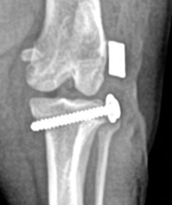
Advertisement
Advertisement
