

News|Articles|August 2, 2011
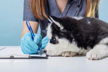
Image Quiz: Ophthalmology-Chronic tears in a springer spaniel
Correct answer for Image Quiz: Ophthalmology-Chronic tears in a springer spaniel
Advertisement
Placement of a nasolacrimal catheter is correct!
Advertisement
This photo shows a tube leaving the upper eyelid (skin) medially. It is attached on the skin a few centimeters further dorsally (not shown). The tube connects to the lower puncta and follows the nasolacrimal duct; the other end is also attached to the surrounding skin after the tube leaves the opening by the nose.
A nasolacrimal duct obstruction was diagnosed as purulent material was expressed from the lower puncta on palpation at the level of the lacrimal sac and a Jones test result was negative in that eye (no evidence of fluorescein in the ipsilateral naris after five minutes). A negative result on a Jones test does not mean that the nasolacrimal duct is blocked; however, in this case, applying topical anesthesia and flushing the nasolacrimal duct was unsuccessful. General anesthesia helps increase the chances of successfully flushing a blocked nasolacrimal duct, which was the case in this dog. If the blockage is relieved, a bacterial culture should be done on the material that is initially flushed out of the duct, and a nasolacrimal catheter can be placed with the patient under general anesthesia. This catheter is left in place for four to six weeks, especially if the blockage is recurrent. If flushing while the patient is anesthetized is unsuccessful, contrast studies are indicated (dacryocystorhinography) to localize the blockage and potentially formulate a surgical plan.
This case highlights the importance of evaluating ocular surface disease and nasolacrimal duct obstruction in every case with increased facial staining.
Advertisement
Related Content
Advertisement


Latest CME


Advertisement
Advertisement
Trending on dvm360
1
Veterinary Emergency Team in Texas deploys in response to NWS cases
2
Investigational drug shows promise for treating feline obesity
3
USDA approves canine vaccination for Lyme disease and leptospirosis
4
Survey highlights gaps in canine pruritus management expectations
5