

|Articles|November 1, 2009
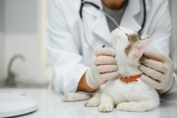
Hypoadrenocorticism in the dog (Proceedings)
Author(s)Jana Gordon, DVM, DACVIM
Hypoadrenocorticism occurs as a result of a deficiency in glucocorticoids and, in most cases, mineralocorticoids.
Advertisement
Hypoadrenocorticism occurs as a result of a deficiency in glucocorticoids and, in most cases, mineralocorticoids. Primary hypoadrenocorticism is the result of destruction of the adrenal cortices. Secondary hypoadrenocorticism occurs as a result of insufficient ACTH secretion from the hypothalamus leading to atrophy of the adrenal cortices and primarily a glucocorticoid deficiency.
Normally neural stimulation of the hypothalamus produces corticotropin-releasing hormone (CRH). Corticotropin-releasing hormone stimulates the release of adrenocorticotropic hormone (ACTH) from the anterior pituitary. ACTH exerts its effects on the adrenal cortex and stimulates the zona fasciculata to release cortisol, the zona glomerulosa to release mineralocorticoids and the zona reticularis to release androgens. ACTH's primarily effects are on cortisol release with minimal effects on mineralocorticoid and androgen production. Cortisol provides a negative feedback to ACTH and CRH. Glucocorticoids are involved in many normal physiologic responses in the body.
Mineralocorticoid release form the zona glomerulosa is primarily mediated by the renin angiotensin aldosterone system and only minimally by ACTH. Renin is released from the juxtaglomerular apparatus of the kidney into circulation as a result of sympathetic stimulation, hypotension, hyponatremia and hypochloremia. Renin then converts circulating angiotensinogen to angiotensin I. Angiotensin converting enzyme in the pulmonary vascular endothelium converts angiotensin I to angiotensin II. Angiotensin II then stimulates aldosterone secretion from the zona glomerulosa. Potassium can also directly stimulate aldosterone production. Aldosterone enhances sodium and chloride retention with hydrogen ion and potassium loss in the renal tubules.
Etiology
The majority of dogs with hypoadrenocorticism have idiopathic atrophy of the adrenal cortices likely secondary to immune-mediated destruction. In humans it is commonly seen as part of an immune-mediated polyglandular syndrome. In dogs polyglandular syndromes involving hypothyroidism, diabetes mellitus and/or hypoparathyroidism have rarely been documented. Hypoadrenocorticism may also occur secondary to cortical destruction as a result of adrenolytic therapy. Chronic administration of glucocorticoids leads to cortical atrophy and rapid withdrawal can lead to hypocortisolism. Infarcts, trauma and neoplasia of the adrenal gland(s) are rare causes of adrenal insufficiency.
Hypoadrenocorticism can occur in any breed but there may be an increased incidence in West Highland white terriers and Great Danes. The disease is inherited as an autosomal recessive trait in Nova Scotia Duck Tolling Retrievers, Portugese Water Dogs and the standard poodle. It is heritable in the bearded collie. Females are affected more frequently than males. Dogs of any age can be affected but the disease is more common in young adult and middle-aged dogs.
History
The history can be variable. The classic history is of waxing and waning lethargy, inappetence, vomiting and diarrhea that often responds to symptomatic treatment. There may be a stressful event that precipitates these episodes. Dogs may also present for weakness, polyuria, polydipsia, muscle wasting, regurgitation and seizures. These signs may be present for weeks to months reflecting the progressive destruction of the adrenal glands. A more acute presentation may also occur in which dogs present in hypovolemic shock. These dogs may or may not have a history of more chronic signs. In acute disease anorexia, vomiting, diarrhea, melena and collapse are more common.
Clinical findings
Dogs may be weak, lethargic, exhibit muscle wasting and have abdominal pain. In a more acute presentation the dog may be laterally recumbent with pale tacky mucous membranes, increased capillary refill, poor pulse quality, bradycardia and hypothermia.
Laboratory findings
The most common finding on the CBC is a mild non-regenerative anemia. In some cases the absence of a stress leukogram (lymphopenia, eosinopenia) is noted due to the absence of cortisol. The classic electrolyte abnormalities are increased potassium, decreased sodium and decreased chloride. These occur due to mineralocorticoid deficiency because of a lack of aldosterone and its effects on the renal tubules. The Na:K ratio has classically been used to aid in the diagnosis of this disease with an ACTH stimulation test recommended in dogs with a Na:K ratio of 27 or less. A study has reported a sensitivity of 89% and specificity of 97% if the ratio of 27 was used. This means that 89% of dogs with hypoadrenocorticism will be indentified as positive using a ratio of 27 (will miss 11% and incorrectly call them negative) and that 97% of dogs without hypoadrenocorticism will be correctly identified as negative (will miss 3% and incorrectly call them positive). Increased BUN and creatinine are primarily prerenal but intestinal hemorrhage can cause increases in BUN. It is possible that these dogs can develop acute renal failure if their prerenal factors (hypovolemia, hypotension) are not addressed in a timely fashion or if they have underlying renal disease or insufficiency but this is uncommon. Liver enzymes may be normal or mildly increased likely due to poor perfusion of the liver. Hypoglycemia may also be seen with cortisol deficiency. A mild to moderate hypercalcemia (total ± ionized) is fairly common and the exact cause unknown but does not appear to be related to parathyroid hormone or parathyroid related protein. Alterations due to increased bone resorption, an increase in intestinal absorption, decreased glomerular filtration rate and increased renal tubular reabsorption may play a role. Mild hypoalbuminemia is also seen frequently and likely due to increased losses in the intestinal tract. Hypocholesterolemia can also occur and may be due to decreased intestinal absorption of fats or mobilization from tissues, both cortisol-dependent functions. A metabolic acidosis occurs secondary to decreased hydrogen ion excretion (aldosterone deficiency), poor tissue perfusion (generates lactic acid) and decreased renal excretion of acids.
Diagnostic imaging
Thoracic radiographs might reveal microcardia, decreased diameter of the caudal vena cava and megaesophagus. On abdominal ultrasound, adrenal glands might be normal or occasionally, small in size.
Electrocardiography
Hyperkalemia can cause several electrocardiographic changes including bradycardia, a spiked T wave, shortening of the Q-T interval, increased duration of the QRS, decreased to absent P wave, prolonged P-R interval and atrial fibrillation or standstill.
Endocrine tests - The ACTH stimulation test
The ACTH stimulation test is the only test available to diagnose this disease. It is important to note that a single baseline cortisol is inadequate for diagnosis and may be low or undetectable in dogs with non-adrenal illness. A study evaluating basal cortisol in dogs with hypoadrenocorticism reported 100% sensitivity and 98% specificity for detecting hypoadrenocorticism when the lower limits of detection of the test are </= 1 mcg/dl but specificity decreased to 78% when the lower limit was </= 2 mcg/dl. This means that all dogs with hypoadrenocorticism have a baseline cortisol < /= 1 mcg/dl but a small number of dogs with non-adrenal illness have a basal cortisol </= 2 mcg/dl. Blood for a baseline cortisol is taken followed by administration of either synthetic ACTH gel given IM followed by a blood draw at 2 hours or synthetic corsynotorpin (250 mcg/dog or 5 mcg/kg) with a second sample obtained an hour later. There should be no to minimal stimulation and cortisol levels are almost always less than 2.0 µg/dl.
Endogenous ACTH
This test can be done to differentiate primary from secondary hypoadrenocorticism. In primary hypoadrenocorticism ACTH is increased because of a lack of negative feedback from cortisol. With secondary hypoadrenocorticism ACTH is low to low normal due to failed secretion from the pituitary. Interestingly, a few dogs with secondary hypoadrenocorticism also have decreased aldosterone levels in the absence of electrolyte abnormalities. Some, but not all, of these dogs go on to develop the more classic electrolyte abnormalities in fact in one study only 1 of 11 dogs with secondary hypoadrenocorticism went on to develop electrolyte abnormalities.
Aldosterone
Aldosterone can also be used as a differentiating test but is more difficult to interpret than endogenous ACTH. Aldosterone is measured prior to and 1 hour after synthetic ACTH is administered as described above. In normal dogs aldosterone should increase significantly. In dogs with primary hypoadrenocorticism, there should be little to no increase in aldosterone and with secondary hypoadrenocorticism, theoretically, there should be an increase in aldosterone. Unfortunately in many cases of secondary hypoadrenocorticism when only ACTH should be low, aldosterone concentrations are also decreased, similar to what is seen with primary hypoadrenocorticism.
Treatment
In hypovolemic shock it is important to address hypotension and hypovolemia. Typically this is initially done with shock rates (80 – 90 ml/kg/hr) of crystalloids until fluid deficits and blood pressure are normalized. After correcting hypovolemia, fluid rates are adjusted to maintain hydration and replace losses. 0.9% Sodium chloride is used initially in dogs with evidence of mineralocorticoid deficiency because of its higher sodium content. If bradycardia and moderate to severe hyperkalemia (> 8.0 mEq/L) are present then 10% calcium gluconate can be administered IV at 0.5 ml/kg over 15 minutes to help maintain a normal membrane potential. A continuous ECG should be used while administering IV calcium. Dextrose (1 g/kg) ± regular insulin (0.2 U/kg) can be given IV to move potassium intracellularly. Alternatively sodium bicarbonate at 2 mEq/kg can be given for translocation and may help improve hyponatremia. Fluid diuresis will aid in elimination of potassium through the kidneys and in mild to moderate hyperkalemia, may be all that is required.
Parenteral glucocorticoids should be given as soon as possible. Glucocorticoids most commonly used are dexamethasone, dexamethasone sodium phosphate, prednisolone sodium succinate and hydrocortisone hemisuccinate. If considering glucocorticoids prior to or during the ACTH stimulation test, dexamethasone formulations and hydrocortisone hemisuccinate are recommended because they reportedly will not interfere with measuring cortisol during the ACTH stimulation test. Initially high doses of glucocorticoids are used more frequently. Dexamethasone sodium phosphate is given at 0.5 mg/kg IV 2 to 3 times a day. This can be followed up by dexamethasone or dexamethasone sodium phosphate at 0.5 mg/kg once daily, hydrocortisone sodium succinate at 0.5 mg/kg/hr CRI or prednisolone sodium succinate at 0.25 to 1.0 mg/kg BID. Parenteral glucocorticoids are given until the dog is no longer vomiting and eating and drinking regularly. At this time dogs can be switched to oral prednisone at 0.5 mg/kg BID then tapered to lowest dose necessary. Maintenance doses of glucocorticoids can be as low as 0.1 mg/kg every other day but it might take several weeks until the dog can be safely weaned to this dose. If utilizing fludrocortisones as a mineralocorticoid, it may be possible to discontinue glucocorticoids because fludrocortisone contains glucocorticoids.
Mineralocorticoids are not necessary in an acute crisis but should be given as soon as a mineralocorticoid deficiency is diagnosed. Options for mineralocorticoid replacement are desoxycorticosterone pivalate (DOCP) or fludrocortisone acetate. DOCP is given at 2.2 mg/kg SC or IM injection every 2 to 6 weeks. Most dogs require injections every 22 to 25 days. Monitoring electrolytes every 2 weeks will help determine dosing intervals. Initially IM administration of DOCP might be preferred due to possibilities of poor perfusion in acute disease. Fludrocortisone is an alternative in a dog that can take oral medications. Fludrocortisone is given at 0.02 mg/kg once to twice daily. DOCP has very little glucocorticoid activity so supplementation with glucocorticoids is necessary. Fludrocortisone can occasionally be administered without glucocorticoids but in both instances glucocorticoids should be given initially and then tapered to the lowest dose necessary to maintain appetite and normal activity levels while minimizing signs of glucocorticoid excess. Sodium and chloride often remain low despite normal potassium. Salt can be supplemented to help normalize sodium and chloride but is usually not necessary long term.
Prognosis
The disease carries a good to excellent prognosis with treatment. Many dogs with atypical hypoadrenocorticism may develop mineralocorticoid deficiency so electrolytes should be checked periodically for the first 4 months.
References
Adamantous S, Boag A. Total and ionized calcium concentrations in dogs with hypoadrenocorticism. Vet Rec 2008. Jul 5;163(1):25-6.
Adler JA, Drobatz KJ, Hess, RS. Abnormalities of serum electrolyte concentrations in dogs with hypoadrenocorticism. J Vet Int Med. 2007. Nov-Dec;21(6):1168-73.
Feldman EC, Nelson RD. Hypoadrenocorticism in Canine and Feline Endocrinology and Reproduction. 3rd ed Feldman EC, Nelson RD, eds. 2004. pp 394 – 435.
Foley C, Bracker K, Drellich S. Hypothalamic-pituitary axis deficiency following traumatic brain injury in a dog. J Vet Emerg Crit Care. 2009 Jun;19(3):269-74.
Gow AJ, Gow DJ, Bell R, et al. Calcium metabolism in eight dogs with hypoadrenocorticism. J Sm Anim Pract. 2009 Aug;50(8):426-30.
Hughes AM, Nelson RW, Famula TR, et al. Clinical features and heritability of hypoadrenocorticism in Nova Scotia Duck Tolling Retrievers: 25 cases (1994 – 2006). 2007 Aug 1;231(3):407-12.
Labelle P, De Cock HP. Metastatic tumors to the adrenal glands in domestic animals. Vet Pathol 2005 Jan;42(1):52-8.
Lennon EM, Boyle TE, Hutchins RG, et al. Use of basal serum or plasma cortisol concentrations to rule out a diagnosis of hypoadrenocorticism in dogs: 123 cases (2000 – 2005). J Am Vet Med Assoc 2007 Aug 1;231(3):413-6.
Melian C, Stefanacci J, Peterson ME, et al. Radiographic findings in dogs with naturally-occuring primary hypoadrenocorticism. J Amer Anim Hosp 1999 May-Jun;35(3):208-12.
Thompson AL, Scott-Moncrieff, Anderson JD. Comparison of classic hypoadrenocorticism with glucocorticoids-deficient hypoadrenocorticism in dogs: 46 cases (1985-2005). J Am Vet Med Assoc 2007 Apr 15:230(8):1190-4.
Advertisement
Related Content
Advertisement

Latest CME


Advertisement
Advertisement