

|Articles|August 1, 2009
Gross dermatology images educate clients
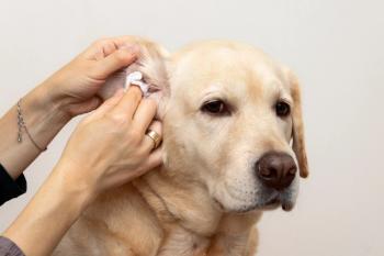
I take digital photos of cytologic exam and skin scraping findings (bacteria, mites) through one eyepiece of the microscope by using the camera's macro setting.
Advertisement
I take digital photos of cytologic exam and skin scraping findings (bacteria, mites) through one eyepiece of the microscope by using the camera's macro setting. Then I show the photos to clients in the exam room. This pictorial representation of the high numbers of "gross" bacteria drives home the necessity for antibiotic therapy and reduces complaints about antibiotic costs.
Dr. Brett Wildermuth, DACVD
Dr. Brett Wildermuth, DACVD
San Diego, Calif.
Advertisement
Related Content
Advertisement


Latest CME


Advertisement
Advertisement
Trending on dvm360
1
FDA-approved sevoflurane inhalant anesthetic for dogs enters veterinary market
2
New World screwworm updates: The latest on government, industry response, new cases
3
‘This is not going to be an isolated incident': Q&A with a parasitologist on the New World screwworm's return to the US
4
New World screwworm confirmed in United States, USDA announces
5