Feline hepatobiliary disease: What's new in diagnosis and therapy? (Proceedings)
Toxic hepatopathy is a direct injury to hepatocytes or other cells in the liver attributable to therapeutic agents or environmental toxins. Cats are particularly sensitive to phenolic toxicity because of limited hepatic glucuronide transferase activity.
Toxic Hepatopathy
Pathogenesis and Etiology – Toxic hepatopathy is a direct injury to hepatocytes or other cells in the liver attributable to therapeutic agents or environmental toxins. Cats are particularly sensitive to phenolic toxicity because of limited hepatic glucuronide transferase activity. The discriminatory eating habits of cats may account for the relatively uncommon occurrence of hepatotoxicity from ingested environmental toxins such as pesticides, household products, and other chemicals. Medical therapies (acetaminophen, acetylsalicylic acid, megesterol, ketoconazole, phenazopyridine, tetracycline, diazepam, griseofulvin) and environmental toxins (pine oil + isopropanol, inorganic arsenicals, thallium, zinc phosphide, white phosphorus, Amanita phalloides, aflatoxin, phenols) may contribute to liver pathology. A severe idiosyncratic hepatotoxicity has been reported with diazepam administration in several groups of cats. Clinical signs in affected cats include anorexia, vomiting, weight loss, ascites, encephalopathy, and death. The histology is characterized by severe central lobular necrosis and mild vacuolation.
Mechanisms of Hepatotoxicity - The liver is an important site of drug toxicity and oxidative stress because of its proximity and relationship to the gastrointestinal tract. Seventy-five to 80% of hepatic blood flow comes directly from the gastrointestinal tract and spleen via the main portal vein. Portal blood flow transports nutrients, bacteria and bacterial antigens, drugs, and xenobiotic agents absorbed from the gut to the liver in more concentrated form. Drug-metabolizing enzymes detoxify many xenobiotics but activate the toxicity of others. Hepatic parenchymal and non-parenchymal cells may all contribute to the pathogenesis of hepatic toxicity. The major mechanisms of hepatotoxicity include: Bile Acid-Induced Hepatocyte Apoptosis, Cytochrome P4502E1-Dependent Toxicity, Peroxynitrite-induced Hepatocyte Toxicity, Adhesion Molecules and Oxidant Stress in Inflammatory Liver Injury, Microvesicular and Nonalcoholic Steatosis.
Diagnosis of Hepatotoxicity – Clinical evidence includes supportive history, normal liver size to mild generalized hepatomegaly, elevated serum liver enzyme activities (predominantly ALT and AST), hypoalbuminemia and hypocholesterolemia, and recovery or death depending upon severity and magnitude of exposure. There are no pathognomonic histologic changes in the liver, although necrosis with minimal inflammation and lipid accumulation are considered classic findings.
Treatment of Hepatotoxicity – Few hepatotoxins have specific antidotes, and recovery relies almost exclusively on symptomatic and supportive therapy. If recognized, acetaminophen toxicity may be treated with acetylcysteine (sulfhydryl group donor), ranitidine or cimetidine (cytochrome P450 enzyme inhibition), ascorbic acid (anti-oxidant), and androstanol (consititutive androstane receptor [CAR] inhibition).
Hepatic Lipidosis
Pathogenesis and Etiology – Feline hepatic lipidosis is now a well-recognized syndrome characterized by intracellular accumulation of lipid with clinicopathologic findings consistent with intrahepatic cholestasis. The precise incidence of the syndrome is unknown but pathology surveys have revealed 5% of animals affected with this lesion. While some cases result from diabetes mellitus, the majority of cases are felt to result from the nutritional and biochemical peculiarities of the cat. It has been suggested, for example, that the cat is not very capable of regulating intermediary metabolism during starvation. Although the biochemistry of this lesion has not been completely worked out, there are several biochemical and nutritional peculiarities that predispose the cat to this syndrome. Some of the known biochemical peculiarities of the cat are: essentiality of dietary arginine; low levels of hepatic ornithine; high dietary protein requirements; lack of hepatic enzymatic adaptation to low dietary levels of protein; relative insufficiency of intestinal pyrroline-5-carboxylate synthase activity; relative insufficiency of intestinal and hepatic glutamate reductase; relative insufficiency of intestinal ornithine transcarbamylase; peculiarities in lipoprotein metabolism; and, differences in orotic acid metabolism.
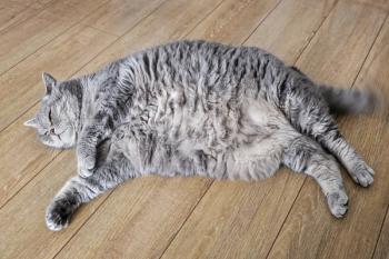
Clinical Features – Most studies suggest that there are no breed, sex, or age predilections. A recent retrospective study by Center and her colleagues suggests that female and middle-age cats are at greater risk for the illness. Obesity may be a predisposing factor, although the syndrome readily develops in fit animals. It has been suggested that obesity followed by a period of anorexia and weight loss are particularly at risk. Cats affected with this syndrome are often presented with a complaint of anorexia, often of several weeks duration. These cats are also commonly presented with jaundice. Other reported clinical signs include vomiting, weakness, weight loss, and diarrhea. Physical examination often reveals dehydration, cachexia, jaundice, and hepatomegaly. All of these findings are also reported in cats with acute pancreatitis and other hepatobiliary disease.
Diagnosis – Hyperechoic changes in the hepatic parenchyma at ultraonography have been cited as a pathognomonic finding, but these changes may be seen in other feline hepatic disorders. Diagnosis should be substantiated by aspiration cytology, or better still, tissue biopsy (percutaneous, trans-abdominal ultrasound guidance, laparoscopy, or open laparotomy). Aspiration cytology has weak sensitivity and specificity, and may miss other diagnoses.
Therapy – Nutritional support is the cornerstone of therapy of this disorder. Most studies suggest that enteral feeding (by "forced" or encouraged feeding, pharyngostomy, gastrostomy, or enterostomy feeding tube) of commercially available cat foods will effect recovery in 90-95% of affected animals. Biourge and his colleagues have characterized some of the metabolic changes that take place during fasting in obese cats. They have been particularly interested in the effects of protein, lipid, or carbohydrate supplementation on hepatic lipid accumulation during rapid weight loss in obese cats. They found that small amounts of protein administered to obese cats during fasting significantly reduced accumulation of lipids in the liver, prevented increases in alkaline phosphatase activity, eliminated negative nitrogen balance, and appeared to minimize muscle catabolism. Carbohydrate supplementation reduced hepatic lipid accumulation, but metabolic abnormalities still developed. Lipid supplementation alone did not ameliorate hepatic lipidosis and even resulted in more severe lipid accumulation than under conditions of fasting alone. The use of benzodiazepine agonists (e.g. diazepam, oxazepam, elfazepam) and 5-HT2 agonists (e.g., cyproheptadine) as appetite stimulants has been encouraged in anorexic cats. These compounds particularly the benzodiazepine agonists, should be used with caution as they may exacerbate pre-existing hepatic encephalopathy. Benzodiazepine agonists have been shown to worsen hepatoencephalopathy in other animal species through activation of the neuronal benzodiazepine/GABA receptor-chloride channel complex.
Feline Cholangitis
Pathogenesis and Etiology – This syndrome has been classified in three different ways:
University of Minnesota Classification (Doug Weisse; 1996) – Lymphocytic portal hepatitis and suppurative cholangitis. This classification system implies that there are two different inflammatory conditions involving the feline liver: inflammatory liver disease (lymphocytic portal hepatitis) and inflammatory biliary tract disease (suppurative cholangitis). Limitations of this classification system – It fails to recognize that acute (i.e., suppurative) cholangitis can progress to more chronic forms (i.e., lymphocytic) of the disease. This system also implies that there is suppuration, which, in fact, is rarely seen. Neutrophilic infiltrates do occur, but rarely does it progress to suppuration. Finally, it's not entirely clear whether lymphocytic portal hepatitis is a distinct clinical entity or just a histologic lesion.
WSAVA International Liver Standardization Group Classification (Multi-Institutional Group; 2002) – Neutrophilic cholangitis, lymphocytic cholangitis, lymphocytic portal hepatitis. This classification system implies that there are acute (neutrophilic) and chronic (lymphocytic) forms of cholangitis, and that there may be a separate form of portal hepatitis in cats. Limitations of this classification system – We still don't know if lymphocytic portal hepatitis is a disease or a histologic lesion.
Neutrophilic cholangitis – This disorder has been seen primarily in young to middle-aged male cats with clinical signs of acute vomiting, diarrhea, anorexia, and lethargy. Physical examination findings often reveal fever, icterus, abdominal pain, and hepatomegaly (<50% of cases). Laboratory findings frequently reveal mild to moderate leukocytosis with mild to moderate elevations in ALT, AST, GGT, and ALP. Based on recent studies, cats affected with this form of cholangitis often have related disease, e.g., pancreatitis and inflammatory bowel disease. The diagnosis of suppurative cholangiohepatitis is achieved by serum liver enzymology; ultrasonographic characterization of the liver parenchyma; culture - bile, gallbladder, cholelith, liver; Gram staining; and, biopsy of the liver and/or extrahepatic biliary system. Common bacterial isolates in affected cases include E. coli, Clostridia, Bacteroides, Actinomyces, α-Strep. The treatment of this syndrome has included appropriate antibiotic based on culture and sensitivity, cholelith removal where appropriate, bile duct decompression if necessary, fluid and electrolyte maintenance, and ursodeoxycholate therapy (10-15 mg/kg P.O. SID).
Lymphocytic cholangitis – Chronic lymphocytic cholangitis is characterized by a mixed inflammatory response (equal numbers of lymphocytes or plasma cells and neutrophils) within portal areas and bile ducts. Other features of chronicity include marked bile duct proliferation, bridging fibrosis, and pseudolobule formation. Chronic cholangiohepatitis may progress to progressive biliary cirrhosis and the death of the patient. Lymphocytic cholangitis may represent a persistent bacterial infection or an immune-mediated response may result in a chronic self-perpetuating disorder. Clinical signs are usually of a chronic, intermittent or persistent nature. With chronic cholangiohepatitis, a long-standing history over a period of weeks or months is more likely. Vomiting, icterus, hepatomegaly and ascites are common findings. Hepatic encephalopathy and excessive bleeding are uncommon unless severe end-stage liver disease is present. The best treatments for this syndrome are not clearly understood. It has been suggested that many cats require multi-component therapy, e.g., glucocorticoids - 1-2 mg/kg PO SID; metronidazole - 7.5 mg/kg PO BID; ursodeoxycholate 10-15 mg/kg PO SID; vitamin K1 - 1.5-5 mg Q 2-3 weeks; dietary manipulation for presumed I.B.D.; and, immune modulation with azathioprine or chlorambucil.
Lymphocytic portal hepatitis – A retrospective review of liver biopsies of cats with inflammatory liver disease identified a subset of cats with lymphocytic portal infiltrates which had histopathologic features distinct from cats with acute or chronic cholangitis. The term lymphocytic portal hepatitis has been proposed for this disorder. As opposed to findings in cholangitis, there is a lack of neutrophilic inflammation, bile duct involvement, infiltration of inflammatory cells into hepatic parenchyma, or periportal necrosis. Lymphocytic portal hepatitis is not associated with inflammatory bowel disease or pancreatitis. Previous reports of progressive lymphocytic cholangitis or lymphocytic cholangitis referred to varying degrees of neutrophilic inflammation and the condition may actually have been a chronic form of cholangitis. Lymphocytic portal hepatitis is a common finding in liver biopsies of older cats, suggesting that it is a common aging change or that a sub-clinical form of disease is prevalent. Lymphocytic portal hepatitis appears to progress slowly with varying degrees of portal fibrosis and bile duct proliferation but no pseudolobule formation. Concurrent hepatic lipidosis is less likely than with cholangitis.
Hepatic Neoplasia
Pathogenesis and Etiology – Primary neoplasms of the feline liver are uncommon. Cholangiocellular carcinoma and hepatocellular carcinoma are the most important of the primary feline liver neoplasms, but they are of very low incidence and therefore minor importance. Metatstatic liver neoplasia are much more important in cats. The most common metastatic tumors to the liver are lymphoma, systemic mast cell disease, hemangiosarcoma, and myeloproliferative disorders.
Clinical Features – Clinical signs are fairly non-specific, but may be similar to clinical signs reported in cats with other liver disorders, for example: lethargy, anorexia, weight loss, and intermittent vomiting. Abdominal effusion, jaundice, and encephalopathy may be seen terminally.
Diagnosis – Laboratory data are also usually non-specific. Elevations in serum liver enzyme activities and abnormalities in bile salt metabolism should be obvious, but they are not remarkably different from cats with other liver disorders. Imaging studies (radiography, ultrasonography) may provide evidence of diffuse hepatomegaly or of discrete tumors involving one or more liver lobes. Definitive diagnosis always requires aspiration cytology, or better yet, tissue biopsy. Aspirates and/or tissue biopsies may be obtained by percutaneous trans-abdominal ultrasound guidance, laparoscopy, or laparotomy techniques.
Therapy – The cell of origin of a metastatic tumor should always be identified, if possible. Chemo- or other therapies may then be selected based on a working knowledge of the biologic basis of the tumor. Focal tumors of the liver may be best managed by hepatic lobe resection.
Extra-Hepatic Bile Duct Obstruction
Pathogenesis and Etiology – Extra-hepatic cholangitis, malignancy, pancreatitis, cholelithasis, and liver flukes (Eurytrema procyonis, Platynosomum concinnum) are the major causes of extra-hepatic biliary obstruction in cats. Progressive cholangitis accounts for over 50% of the cases of hepatic duct obstruction, common bile duct obstruction, and progressive hepatobiliary failure.
Clinical Features – Affected cats have marked persistent hyperbilirubinemia, and marked elevations in serum ALT, AST, ALP, GGT, and serum bile acids. Ultrasonographic evidence of obstruction is obvious, and many cats undergo exploratory laparotomy and biliary decompression.
Diagnosis – As with other feline hepatobiliary disorders, diagnosis of extra-hepatic bile duct obstruction requires careful integration of history, physical examination, laboratory data, and imaging findings.
Prognosis and Therapy – The prognosis for cats with extra-hepatic biliary obstruction, regardless of underlying pathogenesis is guarded to poor, and perioperative morbidity and mortality is high. The majority of cats have a prolonged disease course, and long-term complications include recurring bouts of cholangitis, weight loss, and biliary tract obstruction.
References available upon request
Newsletter
From exam room tips to practice management insights, get trusted veterinary news delivered straight to your inbox—subscribe to dvm360.


