Dental and oral examination: The normal oral cavity of dogs and cats (Proceedings)
Familiarity with the normal structures and physiology of the oral cavity is a powerful tool to help identify what is not normal. Detailed examination of many normal mouths is the best way to acquire expertise.
The mouths of dogs and cats are beautiful things, ideally designed to perform their functions. Consider the canine skull; the facial bones extend far forward from the cranial vault to provide large areas with enough space to fit many teeth strongly anchored in bone. This elongated area simultaneously increases the surface area of the nasal mucosa. Without strong, sharp, healthy teeth an animal in the wild would starve. Their first set of teeth are deciduous; smaller, sharper, more fragile, and fewer in number. As a puppy learns to use its teeth, the deciduous teeth are often fractured as they use these brittle structures as their main interface with, and means of, exploring their environment. So it works well that they are replaced by a second permanent set. By the time the permanent teeth are erupting there is much more space in the alveolar arches to accommodate the larger size and greater number of teeth. Eruption is completed only shortly before full dental arch length is attained, completing the dental arches.
Familiarity with the normal structures and physiology of the oral cavity is a powerful tool to help identify what is not normal. Detailed examination of many normal mouths is the best way to acquire expertise. Quite a few normal structures can appear abnormal, even dangerous the first time they are "discovered". This session will take an in-depth and detailed look at the normal oral cavity of dogs and cats. In the following session this will be carried to the next step by looking at examples of dental and oral pathology.
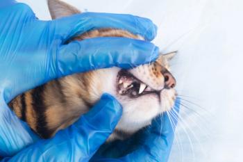
A complete oral examination should be a part of every physical exam. The first step is to check the face both visually and through palpation for symmetry and any evidence of swelling. The lips are lifted and the labial and buccal surfaces of all the teeth are examined. The mouth is opened to examine the dental occlusal surfaces, the palate, and the tongue including the sublingual tissues.
The Teeth
The mature dog has 42 teeth and the cat has 30. This is compared to humans that have 32. In people, many have their 3rd molars ("wisdom teeth") removed leaving them with only 28. Teeth are vital complex structures with a central endodontic system made up of vascular, nervous, and connective tissue (the dental pulp) that occupies the pulp cavity. The pulp cavity is composed of the pulp chamber in the crown and the root canal in the root. The majority of the hard tissue of a tooth in dogs and cats is made of dentin, a combined tissue with a mineralized hard tissue component that is permeated by numerous tubules. The dentin tubules contain cell components (cell processes from the odontoblasts that line the pulp) and fluid. The crown dentin is covered and protected by enamel – a much harder and more highly mineralized material that is impervious to leakage or bacteria when it is intact. The root dentin is covered and protected by cementum, into which the periodontal ligament fibers attach. The tooth is suspended in the alveolus by the periodontal ligament, another physiologically active tissue that contains circulation and sensory feedback. The ligament attaches the cementum of the root to the alveolar bone (compact bone with multiple vascular openings), making the attachment a type of joint called a "gomphosis". In dogs and cats, a very small amount of tooth mobility can be normal for the incisor teeth, but no other teeth should exhibit any mobility. They only become mobile with loss of the supportive tissues.
Enamel is completely formed before a tooth erupts. Once eruption occurs, no more enamel is made. The enamel of dogs is much thinner than that of humans, and cat enamel is thinner yet. Even very small fractures can expose the dentin. When the impervious enamel loses integrity, either through trauma (fractures, abrasion, attrition) or developmental abnormalities, the exposed dentinal tubules can cause dental sensitivity and can allow the ingress of bacteria to the pulp. Dentin can also be exposed by damage to the cementum on the root, for example due to periodontitis or gingival recession.
The pulp is living connective tissue that in health is protected inside the tooth. Its main function is to form dentin. In contrast to enamel, the dentin continues to be formed throughout life as long as the tooth remains vital. A very young puppy or kitten has very thin dentin, and the tooth has the appearance of a very large pulp with an eggshell-thin hard tissue component. A geriatric dog or cat tooth is composed nearly completely of hard tissue with a very thin central root canal space. In puppies the circulation enters the root tip through a large foramen. This apex "closes" before a year of age after which the circulation enters through multiple canaliculi that form an apical delta. There are also some lateral canals and furcation canals that enter through the sides of the root or in the furcation area. All these vascular entry sites can become ports of exit for inflammatory mediators or bacteria when a pulp becomes infected.
Alveolar Bone
The teeth are supported by alveolar bone that forms an arch of bone on the ventral margins of the maxillary and incisive bones and the bulk of the mandibles. The external surfaces of the alveolar processes are cortical bone. The dental alveoli ("sockets") are also lined by compact bone that appears as a white line on radiographs called the lamina dura. Trabecular bone occupies the space between these compact bony layers. The maxillary canine tooth roots extend dorsally in the vertical process of the maxilla to lie lateral to the nasal cavity, while the root tips of the maxillary molars extend to the area of the sphenopalatine fossa in the orbit. The anatomical positions of the root tips determines where infection of endodontic origin will damage adjacent tissues.
Soft Tissues
The alveolar bone is covered along the margin and around the teeth by attached gingiva. The gingiva receives its blood supply from the adjacent mucosal vessels, from the marginal vessels in the sulcus, and from the periosteal circulation. It is tightly attached to the bone and immovable. At the mucogingival line, the tissue transitions to a loosely attached alveolar mucosa. The mucosa receives its blood supply from vertically-oriented vessels. The vascular supplies need to be considered during mucogingival surgical procedures. This is less critical for gingiva with its triple vascular supply, but becomes important for mucosa with its more limited supply.
An incisive papilla is located just behind the maxillary incisors on the palate. It can be mistaken for a pathological enlargement. Palatal rugae cross the palate horizontally relatively symmetrically. They can be more arched in dolicocephalic breeds, and can develop deep crevices that entrap hair and debris on some brachycephalic breeds.
The tongue is attached at the base and has a free apex with a ventral mucosal fold called the frenulum. Intrinsic tongue muscles change the shape of the tongue while extrinsic muscles reposition it. The most important function of the tongue is for eating and drinking, although it has numerous secondary functions such as grooming in cats and thermoregulation in dogs.
Anatomical Nomenclature
When describing the facial bones and general orofacial structures, the terms "dorsal" and "ventral" are used. However, due to peculiarities of the oral cavity, different anatomical terms are used to describe surfaces and directions on the individual teeth. When considering any single tooth, the surface toward the outside of the mouth is considered the labial surface for rostral teeth and the buccal surface for caudal teeth. It can also be called the "facial" surface. The tooth surface towards the inside of the mouth is the lingual surface. It can also be called the palatal surface on maxillary teeth. The sides of the teeth are referred to as "mesial" for the surface toward the midline of the arch, and "distal" for the surface away from the midline of the arch. This means that for incisors, "mesial" is the medial side and "distal" is the lateral side. But for canines, premolars, and molars, "mesial" is the rostral side and "distal" is the caudal side. The mesial and distal surfaces are closest to their neighboring teeth, so are called "proximal" surfaces. Thus the spaces between the teeth are the "interproximal spaces".
The vertical direction of individual teeth is divided into "apical" towards the root and "coronal" towards he crown. Therefore the term "apical" refers to the dorsal direction for maxillary teeth and ventral for mandibular teeth.
Occlusion
Normal incisor tooth occlusion in dogs is somewhat similar to humans. They meet in a normal scissors bite with the maxillary incisors positioned labial to (in front of) the mandibular incisors. The cusp tips of the mandibular incisors touch the palatal surface of the maxillary incisors on the cingulum. The lower canine is positioned midway between the upper 3rd incisor and canine, and the upper canine is positioned just behind the lower canine. The upper 1st premolar tooth is centered in the interproximal space between the lower 1st and 2nd premolars. This relationship continues caudally, with the teeth of the upper jaw in the center of the space behind the corresponding lower teeth in a saw-tooth pattern. Deviations from this are considered malocclusion.
Dental Charts and Numbering
Use of a numerical dental "shorthand" to identify teeth facilitates recording findings and treatments on dental charts. A common numbering system used in the United States for human mouths, with eight teeth per quadrant, starts at the upper right 3rd molar as number 1, and then are numbered sequentially along the arch around the front making the central incisor tooth number 8, and on around to the upper left last tooth (number 16). The left lower third molar starts at number 17 and on through 24 for the lower left central incisor, and continued around to the lower right quadrant numbered 25 through 32. This system works very poorly for non-primate animals, since different species have different numbers of teeth. Of the many numbering systems used for dogs and cats, the most commonly used is the Modified Triadan System that is very similar to the numbering system used in most other countries for human mouths and recommended by the International Dental Federation. Both systems number the quadrants in a clockwise fashion. As you look at your patient, the quadrant on your upper left (patient's upper right) is 1, then moving clockwise to the upper right (patient's upper left) is 2, patient's lower left is 3, and patient's lower right is 4. In the human system, the teeth within each quadrant are then numbered 1 through 8. So a human's upper right 1st incisor would be number 11 (quadrant 1, tooth 1) and a right maxillary 3rd molar would be 18 (quadrant 1, tooth 8), while the same teeth on the person's left side would be numbers 21 and 28.. Many of our patients have over 9 teeth per quadrant, so we modify this by numbering the teeth 01 through 12 (for the archetypal carnivore). So a dog's upper right 1st incisor is #101. The upper right canine is #104. Upper right 4th premolar is #108, left is #208, lower left 1st molar is 309 etc.
The numbering gets a little trickier for cats. By convention 4th premolar teeth (commonly the last premolar in carnivores) is always a number 8, and the 1st molar is a 9. Cats are missing their maxillary 1st premolars and their mandibular 1st and 2nd premolars, so on a cat's right side the numbers jump from 104 to 106 (no 105) on the top and from 404 to 407 (no 405 or 406) on the bottom. They also have only one molar in each quadrant, considered to be the first molar with the 2nd and 3rd absent. In both dogs and cats the maxillary 4th premolars and the mandibular 1st molars are the largest caudal teeth in the quadrants and are called "carnassial" teeth.
Summary
The oral cavity is a rich and diverse environment that is critically important to the life and health of our patients yet at the same time prone to many types of pathology. It is important to be familiar with all the normal structures and relationships in the mouth so that you will be able to readily recognize abnormalities when they are present.
Newsletter
From exam room tips to practice management insights, get trusted veterinary news delivered straight to your inbox—subscribe to dvm360.


