Zoonotic parasitic infections contracted from dogs and cats: How frequent are they?
Veterinarians in practice are on the front lines in preventing transmission of pet-associated zoonotic parasite infections because of their knowledge of the potential risks and through their contact with pet owners.
The popularity of dogs and cats as pets in the United States continues to increase. Recent surveys estimated that there were 73 million owned dogs and 90 million owned cats in the United States, and almost 60% of U.S. households owned a pet.1 The highest rates of dog and cat ownership occur in households of families with young children. Dogs and cats are hosts to many intestinal parasites that may be transmitted to humans through direct contact with infected pets or exposure to environments contaminated with infected animals' feces (Table 1). Children are often at greatest risk of zoonotic infections because of their play habits and affection for pets.

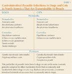
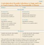
Table 1: Gastrointestinal Parasitic Infections in Dogs and Cats in North America That Are Transmissible to People
Veterinarians in practice are on the front lines in preventing transmission of pet-associated zoonotic parasite infections because of their knowledge of the potential risks and through their contact with pet owners. Practicing veterinarians' services should include preventive treatments to eliminate parasites as well as advice to owners on minimizing the risk of zoonotic transmission. This article summarizes data on the modes of transmission of potentially zoonotic intestinal parasites of dogs and cats and the available data on the frequency at which they infect and cause disease in humans in the United States.*
HELMINTH INFECTIONS
Potentially zoonotic gastrointestinal parasites of dogs and cats include the maternally transmitted intestinal roundworms and hookworms whose infective stages may contaminate and persist in the peridomestic environment (i.e. in proximity to humans).


Peter M. Schantz, VMD, PhD
Ascarids
Infection of humans by Toxocara canis and Toxocara cati, the common roundworms of dogs and cats, respectively, cause larva migrans syndromes (visceral and ocular larva migrans and covert toxocariasis) in humans who accidentally ingest infective eggs from contaminated environments.2 Toxocariasis ranks among the most common of all zoonotic infections; the results of numerous published surveys document seroprevalences in humans ranging from 1% to 20%, depending on the age, socioeconomic status, and pet ownership status of the tested populations.3-5 Toxocaral larva migrans, or infection by the common ascarid worms of dogs and cats, is arguably the most common zoonotic infection associated with pets in the United States and other industrialized countries.4 It has been estimated that every year in the United States this infection causes hundreds of cases of unilateral blindness and uncountable numbers of less permanent forms of illness in children.3,4 The severity and type of disease in humans produced by Toxocara species infection depend on how many larvae are ingested, the frequency of reinfection, and other factors still poorly understood. Most human infections with Toxocara larvae are well-tolerated, even asymptomatic; however, a proportion of infected people develop larva migrans syndromes that may be systemic or confined to the eye. When a larva invades the eye, it almost always leaves the individual partially or totally blind in that eye. Based on data obtained from the Centers for Disease Control and Prevention (CDC) serologic diagnostic reference service, an estimated minimum of 750 cases of ocular larva migrans are diagnosed by physicians every year in the United States.4 Chart review of patients with a diagnosis of uveitis at the University of California Medical Center determined that ocular larva migrans accounted for 1% of cases of uveitis seen between 1977 and 1996.5 All cases were associated with vision loss in the affected eye. The number of cases of toxocaral visceral larva migrans syndrome is much greater; however, estimates of these are quite imprecise.4
Epidemiologic investigations have consistently determined that the principal risk factor for infection was the presence of a household dog, particularly a pup, in a patient's household within six months of onset of illness.2,3,6 When this condition is combined with pica, especially dirt eating, the statistical association becomes very strong.
Hookworms
Infections with the hookworms Ancylostoma braziliense and Ancylostoma caninum remain common in dogs and cats, with the highest prevalences in the southern United States, mainly in coastal areas from southern New Jersey to the Florida Keys and westward along the Gulf of Mexico to Texas. Infections in people are acquired from contact with moist or wet sand or loam soil containing filariform larvae of hookworms generated from the feces of dogs and cats, usually in unprotected sandboxes, on bathing beaches, and under houses where workers lie prone while repairing leaking water pipes. Larval invasion of skin in humans produces pruritic papules. In two or three days, these papules become serpiginous tunnels in the epidermis caused by inflammation resulting from intradermal migration of larvae (cutaneous larva migrans).7 Without treatment, migration may continue for several weeks or months before the immune system kills the larvae. Zoonotic hookworm infection may also be acquired through ingestion of the larvae in soil or in tissues of paratenic hosts. Infection in humans acquired by these latter routes, especially A. caninum, may occasionally lead to enteric localizations of zoonotic hookworms, causing eosinophilic enteritis.8 Although eosinophilic enteritis has been diagnosed with relative frequency in Australia where it was first noted, it has rarely been diagnosed in the United States.9 The eosinophilic enteritis syndrome requires clinical experience and technical sophistication to diagnose and may occur more frequently than currently recognized and documented.
Tapeworms
Dipylidium caninum. Zoonotic tapeworm infections associated with dogs and cats include the flea tapeworm, Dipylidium caninum. Infection is acquired when a person, usually a young child, accidentally ingests a flea carrying the larval stage of the tapeworm. Dipylidium caninum infection can lead to diarrhea and pruritus in infected humans. This infection rarely causes serious symptoms; however, the stress associated with seeing tapeworm segments in a child's stool or diapers can be considerable.10
Echinococcus species. Echinococcus species of dogs may infect humans with larval stages that cause cystic or tumorous growths in the liver and other visceral organs. Cystic echinococcosis, or hydatid disease, is caused by infection with larval stage Echinococcus granulosus. Cases of cystic echinococcosis acquired in the northernmost regions of North America, especially Canada and Alaska, are caused by the northern sylvatic genotype maintained in cycles involving wolves, dogs, moose, caribou, and other cervids.11 The practice of feeding the viscera of moose and caribou to working and pet dogs leads to infection in dogs and subsequent exposure to humans. Infection continues to be relatively commonly diagnosed in most Canadian provinces. A review of hospital records in Edmonton, Alberta, noted 42 cases diagnosed and treated between 1991 and 2001.12
Foci of local transmission involving a variety of domestic intermediate hosts have been described in various regions of the United States.11 Distinct foci of E. granulosus transmission were noted in the 1970s in western states including California, Utah, New Mexico, and Arizona. Epidemiologic investigations revealed that transmission was associated with unique cultural practices involving home slaughter of sheep and the access of dogs to discarded viscera of these hosts. Human populations at risk in these settings were transhumant sheep ranchers, including Basque-Americans in California, Mormons in central Utah, and Navajo and Zuni Indians in New Mexico and Arizona. Active transmission appears to have been eliminated in some of those foci; however, local hospital records indicate that an average of one to four cases continue to be diagnosed each year among Native American communities in Arizona and New Mexico.11
Echinococcus multilocularis, the cause of the alveolar form of human hydatid disease, is an emerging zoonotic parasite in the United States. The life cycle of E. multilocularis involves foxes and coyotes and their rodent prey in ecosystems generally separate from that of humans. However, there is ecologic overlap with humans because fox and coyote populations have increasingly encroached upon suburban and urban areas of many regions, and domestic dogs or cats may become infected when they eat infected wild rodents.11 Infections in domestic pets increase the risk of human exposure. Humans may acquire infection when they accidentally ingest eggs by direct or indirect fecal-oral contamination from infected definitive hosts. Human alveolar echinococcosis in North America has been mainly confined to certain Eskimo populations in northern coastal Alaska in which annual diagnostic incidence rates during the 1970s and 1980s were among the highest ever reported for this infection (7 to 98 per 100,000 population).13 A control intervention in endemic Alaskan villages initiated in 1990 involving education, improved housing, and preventive treatments of dogs has greatly reduced or eliminated transmission to humans.14 No new cases have been diagnosed in humans since that time.14
This tapeworm also occurs in a large area of central North America, and its geographic range and prevalence may be increasing. Before 1964, there were no reports of E. multilocularis in North America south of the Arctic tundra zone, but, in that year, it was reported in foxes and rodents in North Dakota.15 Subsequent surveys revealed that the cestode was enzootic in cycles involving red foxes, coyotes, and deer mice in North and South Dakota, Minnesota, Montana, Iowa, Wyoming, Nebraska, Wisconsin, and Illinois.16,17 The most recent surveys have extended the range eastward to Indiana, Michigan, and Ohio.18,19 Prevalence of infection in foxes and coyotes in the northern Great Plains (25% to 90%) is as high as in any region in the world. To date only two persons are known to have acquired their infections in the endemic region in central North America—a 54-year-old man from Manitoba, Canada, and a 60-year-old woman from Minnesota20 —however, the potential exists for a more serious public health problem as domestic dogs and cats become involved in the life cycle.
Taenia species. Coenurosis is an infection by larval forms of several related tapeworms of the genus Taenia (formerly designated Multiceps). The coenurus is a fluid-filled cyst that measures from a few millimeters to 2 cm or more in diameter. Dogs and other canids (wolves, coyotes, foxes) are the final hosts of Taenia tapeworms. Taenia serialis, the only coenurid-forming cestode currently present in North America, uses rodents or hares as intermediate hosts, and the coenuri are typically found in the intermuscular fascia and subcutaneous tissues. Humans become infected when they accidentally ingest tapeworm eggs in the feces of infected canids. The symptoms of coenurosis are due to the physical presence of the cyst and depend on the site of localization. In North America, fewer than 10 autochthonous (locally acquired) human cases have been documented; three involved the central nervous system or the eye, and the others involved intramuscular or subcutaneous localization.21
PROTOZOA INFECTIONS
Toxoplasma gondii
Toxoplasma gondii is a coccidian parasite widely dispersed in nature. Cats are the definitive hosts for this protozoan, which they acquire when they eat infected intermediate hosts (rodents and many other mammals) or ingest oocysts excreted in the stools of other infected cats. Infected cats are important in the epidemiology and public health importance of toxoplasmosis because they excrete and widely disperse the environmentally resistant oocysts.22 Numerous herbivorous and omnivorous animals become infected when they ingest infective oocysts in soil or contaminated food.
Humans become infected by ingesting food and water contaminated with oocysts shed in the feces of infected cats, by eating undercooked meat from infected animals, or in utero (by congenital transmission from infected mothers). Rarely, humans become infected through blood transfusion or organ transplantation. Recent serosurveys of the U.S. population have documented antibodies (evidence of current or past infection) in about 23% of the U.S. population.23 Ingestion of oocysts shed in the feces of infected cats is believed to directly account for up to 50% of human cases in the United States. Clinical disease caused by toxoplasmosis is generally mild following primary infection of immunocompetent people. Self-limiting fever, malaise, and lymphadenopathy are the most common clinical abnormalities, and most infected people never realize when their first T. gondii infection occurred. However, acute infections acquired by pregnant women can be transmitted to the fetus and cause severe illness (e.g. mental retardation, blindness, epilepsy) and death. According to a 1999 report by the CDC, an estimated 400 to 4,000 cases of congenital toxoplasmosis occur each year in the United States.24,25 Another permanent manifestation of toxoplasmosis is ocular disease, which is estimated to occur in up to 12,000 people per year in the United States. Toxoplasmosis can cause more severe or fatal illness in people who are immunosuppressed (people with human immunodeficiency virus [HIV], transplant recipients).
Giardia species
Giardia duodenalis (synonyms Giardialamblia, Giardia intestinalis) is a protozoan parasite that infects the intestinal tract of many animal species including humans. Motile trophozoite stages occur in the intestines, and environmentally resistant cysts are passed in the feces of infected animals, which are immediately infective if ingested by other susceptible hosts. In all hosts, G. duodenalis can cause acute gastrointestinal signs as well as chronic disease, including chronic malabsorptive and allergic manifestations and childhood failure to thrive.26 Transmission of infection occurs by fecal-oral routes either by direct contact or by ingestion of contaminated food or water. Giardia species infections are common in dogs and cats throughout North America; however, prevalences are often underestimated because the parasite detection methods commonly used in practice have low sensitivity.27,28 Giardia species have long been considered zoonotic because morphologically similar organisms infect humans and a variety of mammals and birds.29 However, evidence of giardiasis being directly transmitted from one host species to an immunocompetent host of another species is limited. Although variants of Giardia species in human, canine, and feline hosts lack differentiating morphologic characters, the application of molecular tools (e.g. PCR) has revealed genetic differences in isolates from different hosts such that it has become clear that the genotypes commonly infecting dogs and cats are not those commonly infecting humans.29,30 Most confirmed infections in humans with Giardia species acquired from dogs or cats have been reported in individuals with recognized immunodeficiency disease (e.g. HIV infection).29
Cryptosporidium species
Protozoan parasites belonging to the genus Cryptosporidium are ubiquitous and among the most common nonbacterial causes of diarrhea in a wide range of vertebrates, including humans. Cryptosporidium species are transmitted via the fecal-oral route by environmentally resistant cysts that are shed in the feces, contaminating soil and water, and, thus, providing multiple routes into the food chain.30 In an immunocompetent host, cryptosporidiosis of the intestinal tract may be asymptomatic or lead to self-limiting diarrhea, but in an immunocompromised host, it can be life-threatening.31 Cryptosporidium species have been reported in numerous mammals and, like Giardia species, appear to have evolved with their respective hosts such that they do not readily cross-infect and develop in hosts of other species. The application of molecular tools has revealed that Cryptosporidium species are a phenotypically and genotypically heterogeneous assemblage of species and genotypes that are morphologically similar.30,31 In humans, the most commonly detected species are the anthroponotic Cryptosporidiumhominis and the zoonotic Cryptosporidiumparvum (cattle). Both Cryptosporidiumcanis and Cryptosporidiumfelis, whose natural hosts are dogs and cats, respectively, have also been demonstrated in infected humans suffering diarrhea.32 Young children and immunocompromised individuals are at greatest risk. Information regarding the role of pets in zoonotic transmission of Cryptosporidium species in immunocompetent humans is insufficient. While it is clear that most outbreaks and individual cases of cryptosporidiosis in humans are related to the contamination of water, food, or fomites with organisms of human or cattle origin, it is also clear that inter-species transmission from dogs or cats to humans can occur in certain situations, especially among very young children or immunodeficient individuals.30
CONCLUSION
Dogs and cats are infected with a number of helminths and protozoa that can infect and sometimes cause life-threatening illness in humans. Awareness of these infections and their zoonotic potential is essential for practicing veterinarians in order to diagnose and treat the infections in pets as well as to provide preventive advice to pet owners.
*None of the zoonotic parasitic infections acquired from dogs and cats are reportable diseases in the United States; consequently, no systematically collected data on the frequency of these zoonotic parasitic infections exist.
Peter M. Schantz, VMD, PhD
Division of Parasitic Diseases
National Center for Zoonotic, Vectorborne and Enteric Diseases
Centers for Disease Control and Prevention
Atlanta, GA 30333
REFERENCES
1. American Veterinary Medical Association (AVMA). U.S. pet ownership & demographics sourcebook. Schaumburg, Ill: American Veterinary Medical Association, 2004:10.
2. Schantz PM. Larva migrans syndromes caused by Toxocara species and other helminths. In: Gorbach SL, Bartlett JG, Blacklow NR, eds. Infectious diseases. 3rd ed. Philadelphia, Pa: WB Saunders Co, 2004;1529-1535.
3. Glickman LT, Schantz PM. Epidemiology and pathogenesis of zoonotic toxocariasis. Epidemiol Rev 1982;3:230-250.
4. Schantz PM. Toxocara larva migrans now. Am J Trop Med Hyg 1989;41(suppl 3):21-34.
5. Stewart JM, Cubillan LD, Cunningham ET Jr. Prevalence, clinical features, and causes of vision loss among patients with ocular toxocariasis. Retina 2005;25:1005-1013.
6. Schantz PM, Meyer D, Glickman LT. Clinical, serologic, and epidemiologic characteristics of ocular toxocariasis. Am J Trop Med Hyg 1979;28:24-28.
7. Davies HD, Sakuls P, Keystone JS. Creeping eruption. A review of clinical presentation and management of 60 cases presenting to a tropical disease unit. Arch Dermatol 1993;129:588-591.
8. Croese J, Loukas A, Opdebeeck J, et al. Occult enteric infection by Ancylostoma caninum: a previously unrecognized zoonosis. Gastroenterology 1994;106:3-12.
9. Khoshoo V, Schantz PM, Craver R, et al. Dog hookworm: a cause of eosinophilic enterocolitis in humans. J Pediatr Gastroenterol Nutr 1994;19:448-452.
10. Molina CP, Ogburn J, Odegboyega P. Infection by Dipylidium caninum in an infant. Arch Path Lab Med 2003;127:e157-159.
11. Moro P, Schantz PM. Cystic echinococcosis in the Americas. Parasitol Int 2006;55(suppl):S181-S186.
12. Somily A, Robinson JL , Miedzinski LJ, et al. Echinococcal disease in Alberta, Canada: more than a calcified opacity. BMC Infect Dis 2005;5:34-43.
13. Schantz PM, Chai J, Craig PS, et al. Epidemiology and control of hydatid disease. In: Thompson RCA, Lymbery AJ, eds. Echinocococcus and hydatid disease, Wallingford, Oxon, UK:CAB International, 233-331.
14. Rausch RL, Wilson JF, Schantz PM. A programme to reduce the risk of infection by Echinococccus multilocularis; the use of praziquantel to control the cestode in a village in the hyperendemic region of Alaska. Ann Trop Med Parasitol 1990;84:239-250.
15. Leiby PD, Olsen OW. The cestode Echinococcus multilocularis in North America. Science 1964;145:1066.
16. Kritsky DC, Leiby PD. Studies on sylvatic echinococcosis. V. Factors influencing prevalence of Echinococcus multilocularis Leuckart, 1863, in red foxes from North Dakota, 1965-1972. J Parasitol 1978;64:625-634.
17. Storandt ST, Kazacos KR. Echinococcus multilocularis in Wisconsin. J Parasitol 1984;70:844.
18. Storandt ST, Kazacos KR. Echinococcus multilocularis identified in Indiana, Ohio, and East-Central Illlinois. J Parasitol 1993;79:301.
19. Storandt ST, Virchow DR, Dryden MW, et al. Distribution and prevalence of Echincoccus multilocularis in wild predators in Nebraska, Kansas, and Wyoming. J Parasitol 2002;88:420-422.
20. Gamble WG, Segal M, Schantz PM, et al. Alveolar hydatid disease in Minnesota. First human case acquired in the contiguous United States. JAMA 1979;241:904-907.
21. Ing MB, Schantz PM, Turner JA. Human coenurosis in North America: case reports and review. Clin Infect Dis 1998;27:519-523.
22. Dubey JP. Toxoplasma gondii oocyst survival under defined temperatures. J Parasitol 1998;84:862-865.
23. Jones JL, Kruszon-Moran D, Wilson M. Toxoplasma gondii infection in the United States, 1999-2000. Emerg Inf Dis 2003;9:371-380.
24. Guerina NG, Hsu HW, Meissner HC, et al. Neonatal serologic screening and early treatment for congenital Toxoplasma gondii infection. The New England Regional Toxoplasma working group. N Engl J Med 1994;330:1858-1863.
25. Jones JL, Lopez AL, Wilson M, et al. Congenital toxoplasmosis: a review. Obstet Gynecol Surv 2001;56:296-305.
26. Caccio SM, Thompson RC, McLauchlin J, et al. Unraveling Cryptosporidium and Giardia epidemiology. Trends Parasitol 2005;21;430-437.
27. Dryden MW, Payne PA, Smith V. Accurate diagnosis of Giardia spp and proper fecal examination procedures. Vet Ther 2006;7:4-14.
28. Hackett T, Lappin MR. Prevalence of enteric pathogens in dogs of north-central Colorado. J Am Anim Hosp Assoc 2003;39:52-56.
29. Thompson RC. The zoonotic significance and molecular epidemiology of Giardia and giardiasis. Vet Parasitol 2004;126:15-35.
30. Slifko TR, Smith HV, Rose JB. Emerging parasitic zoonoses associated with water and food. Int J Parasitol 2000;30:1379-1393.
31. Xiao L, Fayer R, Ryan U, et al. Cryptosporidium taxonomy: recent advances and implications for public health. Clin Microbiol Rev 2004:17:72-97.
32. Xiao L, Ryan UM. Cryptosporidiosis: an update in molecular epidemiology. Curr Opin Infect Dis 2004;17:483-490.
Newsletter
From exam room tips to practice management insights, get trusted veterinary news delivered straight to your inbox—subscribe to dvm360.


