

|Articles|April 1, 2009
Dermatology for technicians (Proceedings)
Author(s)Paul Bloom, DVM, DACVD, DABVP
When an animal is presented with a chief complaint of skin disease it is essential that you approach the problem in a systematic manner.
Advertisement
When an animal is presented with a chief complaint of skin disease it is essential that you approach the problem in a systematic manner. Not only will this be the most cost effective approach but also it is also less likely that you will misdiagnose the underlying disease.
Obtaining the signalment is the first step in the long, winding road toward diagnosing skin disease. Species, age, breed and sex can help point you in the right directions. A puppy or kitten that is presented for skin disease makes you think of ectoparasites, genetic or congenital abnormalities, fungal infections and nutritional problems. A young adult cat or dog would make you think of allergic skin disease, ectoparasites, autoimmune skin disease and endocrinopathy while a geriatric dog would more likely have neoplasias, endocrinopathies or ectoparasites.
The next step may be the most important one, obtaining a detailed history! The very first thing I tell owners with an animal with skin disease is that many diseases look identical because the skin can only respond in a limited number of ways, regardless of cause. I then go on to ask them if they could they tell me how I cut my hand by just looking at the cut? Of course not. That is because a cut on my hand, looks like a cut on my hand regardless of whether it was caused by a piece of glass, a knife or a piece of wood. This is true for animals with skin disease. Therefore, just like a good detective novel, we need to begin at the start and retrace the "footsteps" looking for clues along the way. Before I go over the questions, it is very important to remember not to '"lead" the witness. Don't ask questions like did you give pills bid for 10 days- it is better to ask how did you use the antibiotic or the green pills? Don't ask was the itching better last summer? It is better to ask was the itching last summer better, worse, the same or, another key choice, you DON'T know, as it is today? If you give them the choice of saying they don't know, they won't feel dumb or guilty (I am a bad pet owner syndrome) in giving that reply- or more importantly won't just give you any answer. Questions that you must ask include:
1. What symptoms have you noticed that concern you. I have owners bring in dogs with the most odiferous, painful ears, yet they are bringing the dog in because it is losing its hair. Or is it because the dog has sores but doesn't mention polyuria and polydypsia;
2. Distribution of the skin disease initially and currently;
3. When did these symptoms first occur? This is another important question, because many owners will only tell you when this current episode of symptoms occurred, not the very first time it occurred;
4. Other than the problem the owner presents the patient for, you must ask all owners if the dog has EVER had any other problems with skin or ear disease.
5. Where does the dog live- indoor, outdoors, both? Describe the environment, especially the outdoor environment.
6. Has the dog been treated for skin or ear disease before? What was the age of onset? The table below is an example of why age of onset of symptoms can be useful in finding the underlying cause.
Common Causes of Canine Pruritus Based on Age of Onset
7. Has there been previous treatment for skin or ear disease? If so, when, with what medication and what was the response to treatment. For example how did the dog's pruritus respond to glucocorticoid therapy?
Responsiveness to Glucocorticoid
8. Is she on heartworm and flea preventative? If so, what product, how often is it administered and is it year round or seasonal? If seasonal, when do they begin and end the treatment? Be careful, some of the spot on products for fleas are only used q 60-90 days, which may be adequate for a non-allergic dog, but certainly is not adequate if they have allergies.
9. Are there any other pets in the household? If so, what kind and are they symptomatic. If they are cats, do they go outside?
10. Are any of the humans in the household showing "new" skin problems? If so, what kind
11. Do they board the dog, take him to obedience school, training or to the groomers? If so, when was the last time?
12. Do they know if the parents of the dog or any siblings have pruritic skin problems? If so, what was done and what was the response?
13. What does the dog eat?
14. If the animal is pruritic, how pruritic is it today? Be sure not to ask is the animal pruritic, but how much does she/he lick, scratch, chew, bite, rub or itch – from 0 (normal dog/cat) to 10 (non stop scratching)? How does that compare to other times?
15. Is today's presentation the best, worse or average since the problem began?
16. How was the progression of the disease? Gradual or sudden?
17. Was there a "rash" first or itching first? Or did they occur simultaneously?
18. Do you notice if the symptoms were better, worse or no different last – then choose a season. I don't like asking if they noticed the dog is better in the winter or worse because again, I think you can lead the answers
After reviewing signalment and thoroughly questioning the owner, the next step is to do a complete dermatologic examination. This is when your veterinarian should get involved. Everything we have discussed prior to this is a technician's responsibility (or should be).
After the examination the veterinarian will have a list of differential diagnoses for the skin problem- NOT just the symptoms. For example, in the case of a pruritic (itchy) dog having a DIAGNOSIS OF PRURITUS IS LIKE MAKING A DIAGNOSIS OF VOMITING. It is a symptom so you need to follow the word pruritus with the phrase "due to". The challenge at this point is to make that "due to" list.
Once the veterinarian has made a list of differential diagnoses (ddx) testing may be required. This is again where the technician should be involved collecting the samples and running the tests. Testing usually will include microscopic examination. In fact microscopic examination is an essential part of a dermatologic evaluation. A great deal of information may be obtained by microscopic evaluation of material collected from the hair, ear canal and/or skin. Skin scrapings, cytological examination and fungal cultures are the most commonly used procedures.
Lastly you should be dispensing the medication that the veterinarian prescribed. You will need to explain what the medications are for and re-emphasize the game plan the veterinarian had outlined.
Laboratory Testing
Skin scrapings are done any time a microscopic ectoparasite is suspected. Equipment needed is a scalpel blade, mineral oil, microscope slides, cover slips & a microscope. Collection technique varies depending on parasite suspected (Demodex, Cheyletiella, Sarcoptes). Always apply oil to the slide & blade
Examples of common skin diseases and appropriate tests and treatment for each disease are listed below.
1. Ectoparasites (fleas, Sarcoptes, Cheyletiella, lice, etc)
a. Depending on the parasite, all animals and the house may need to be treated
b. Demodex canis
I. Demodex canis is a follicular mite. It is considered a normal fauna of skin. It proliferates and causes disease either due to an inherited immune defect (juvenile onset) or due to immune suppression (adult onset).
- There are at least 2 other Demodex mites that affect dogs – D. cornei that is a small bodied surface dwelling mite (like D. gatoi in cats) and the other mite is a large bodied follicular mite- D. injai
II. To properly do a skin scraping for Demodex, choose an area that is alopecic, hyperpigmented or where a papule/pustule is present. Demodex cornei has been associated w/a very greasy dermatitis so be sure to scrape greasy areas too. Clip a small "window" in the haircoat if necessary to get easier access to the skin. Squeeze the skin & then scrape until capillary oozing is evident. It is essential that deep skin scrapings be done!! (capillary oozing).
III. Record the stages present (eggs, larva, nymph and adults) and the quantity. Also note how many of the mites are alive and how many are dead. These results will be important to compare to future skin scrapings as you are monitoring the dog's response to therapy.
IV. Demodex is usually easy to demonstrate, however, you may miss in it Shar Peis or if there is chronic pododemodicosis. There are a few pitfalls to avoid when skin scraping dogs for demodex. These include
- Failing to record location of scrapes;
- Failing to record numbers & stages present;
- Failing to record whether the mites are alive or dead
c. Demodex in cats
I. There are 2 types of Demodex in cats, Demodex cati - a follicular mite, and Demodex gatoi - a surface dwelling mite. Both types are normal fauna of skin.
II. D. cati, like D. canis will cause disease in cats that are immunosuppressed. D. gatoi, in contrast to D. cati and D. canis is potentially CONTAGIOUS to the other cats in the household.
III. To properly do skin scraping you need to take into consideration the normal location in the epidermis of the mite. For D. cati, squeeze the skin & then scrape until capillary oozing is evident just as you do for D. canis. Record results as previously described for the dog. These mites are easy to demonstrate.
IV. For Demodex gatoi, since they are superficial dwelling mites, broad superficial scrapings are essential. Clinical signs associated with feline demodicosis include: erythema, crusting, scaling, alopecia and a variable degree of pruritus (mild to moderate). The distribution of the lesions may involve the ventrum; ears, head, neck or it may be generalized.
d. Scabies
I. Sarcoptes scabeii var canis lives in the superficial epidermis of the dog. You need to take broad based, superficial scrapings when you suspect sarcoptes.
II. Because scabies causes a hypersensitivity reaction, it doesn't take a lot of mites to make the dog intensely pruritic. In fact, there is frequently very small number of mites present on the dog. Taking MULTIPLE scrapings (10-15) is essential and false negatives are common (50%). The areas that you should scrape include the ear margins, elbows, ventral chest, & hocks. Choose a non-excoriated area that has papules &/or crusts.
III. Finding 1 mite or egg is diagnostic!!
IV. This mite can temporarily live on humans (zoonosis)
e. Cheyletiella
I. It is also known as "walking dandruff" since animals with Cheyletiella frequently have significant scaling.
II. This mite is NOT host specific and therefore will live on any of the species of animals in the house (so all household haired pets, including ferrets and rabbits must be treated as does the environment).
III. This mite lives on the surface of the epidermis so you want to be sure to take broad superficial scrapings.
IV. These mites also can be difficult to demonstrate, especially in cats. Other methods that may help demonstrate Cheyletiella include: acetate tape impression; flea combing (actually this is the most reliable technique, but still may miss up to 50% of the cats infested) and fecal flotation.
V. Like canine scabies this mite can temporarily live on humans (zoonosis).
2. Malassezia dermatitis
a. Caused by Malassezia pachydermatis which is a commensal
b. Causes pruritic skin and ear disease- hypersensitivity reaction to the yeast
c. Predisposing causes- allergies, recent steroid use (or a dog w/Cushings disease), high environmental humidity, hypothyroidism, changes in the cutaneous microenvironment (excessive sebum production, accumulation of moisture) and breed
d. Diagnosis
I. History
II. Physical examination
III. Skin cytologies
a) Cytologic examination is another very commonly performed procedure in dermatology. Direct smears is used for identifying Malassezia, bacteria and inflammatory cells involved in skin diseases and otitis externa. A slide is rubbed against the affected again to collect surface material. Once the material is on the slide, the slide is heat fixed with a cigarette lighter. The sample is then stained with a 3 step, modified Wright's stain (Diff- Quik). I don't use new methylene blue, even though it gives more nuclear detail, because it gives less detail of organisms. You then air dry and examine microscopically starting with 10X and progressing to finally 100X (oil immersion)
b) If you find 1 organism every 1-3 fields (400X) OR there is ANY field w/more than 1 Malassezia present- this is abnormal and needs to be treated
e. Treatment.
I. Topical treatment
II. Systemic treatment
III. Find the underlying disease
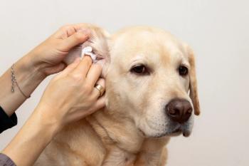
3. Otitis externa is another clinical situation in which microscopic examination is critically important in determining proper management. Treatment decisions are guided by the microscopic findings. To identify mites, take a cotton tipped applicator stick that has been coated with mineral oil, collect debris from the ear canal, place the sample on a microscope slide and then place a cover slip over it. Examine the sample under 10 and 40X magnification.
a. A separate swab for cytological examination is collected to quantitate the numbers & type of bacteria, Malassezia & WBC's present PRIOR to the mineral oil sample. Any time you perform a culture on an ear sample, you should also do cytology. Cytology may identify the presence of Malassezia, which won't be consistently detected on a bacterial culture. Also, there will be instances where the cytology reveals organisms but the culture is negative. This may be due to the presence of anaerobic organisms (they won't grow in a standard, aerobic bacterial culture medium) or fastidious aerobic bacteria.
Treatment for the Allergic Dog
Since allergies are a very common complaint in small animal practice we will review in more details the therapies available. Allergen specific immunotherapy ("allergy shots") is beyond the scope of this presentation.
1. Glucocorticoids are one of the most effective treatments for non-complicated atopy, and are also the class of drugs with the most serious side effects and potential for misuse. They should be used for uncomplicated SEASONAL allergy for SHORT term relief
2. Immunophilin-binding macrolides (cyclosporine (CSA) and tacrolimus)
a. Side effects in dogs are very limited and are primarily GI. Other side effects reported include cutaneous papillomatosis and hyperplastic gingivitis.
b. Drug interactions – the most important is ketaconazole (KCZ). The combination of CSA and KCZ can lower the amount of CSA that needs to be administered.
c. I use this drug in dogs with AD (note that AD has already been diagnosed) when in a moderately to severely pruritic dog:
I. The dog has been on chronic GC at the time I see the dog or;
II. The owners doesn't want to use GC or;
III. While awaiting response to allergen specific immunotherapy and any of the above situations
3. Antihistamines –may be effective in mildly pruritic dogs
4. Essential fatty acids (EFA)- omega 3 (EPA) and 6 fatty acids (GLA and DGLA) may be effective in mildly pruritic dogs especially if used in combination w/antihistamines
Advertisement
Related Content
Advertisement


Latest CME


Advertisement
Advertisement
Trending on dvm360
1
Weekly Vet Report: 9 Screwworm cases (including a dog), new OTC screwworm treatment authorized, & more
2
FDA authorizes first generic drug to combat NWS myiasis in pets
3
ACVIM celebrates award-winning 2026 resident research abstracts
4
Fiber-blend supplement shows promise for gut health in study
5