
|Articles|November 1, 2013
Canine Cushing's Case Files: The ins and outs of detection and treatment-Case file: Libby (Sponsored by Dechra Veterinary Products)
Author(s)Todd M. Archer, DVM, MS, DACVIM
This case illustrates the need to evaluate the patient history and results of routine blood tests to help identify hyperadrenocorticism as a possible differential diagnosis.
Advertisement
Case file: LIBBY
12-year-old spayed female toy poodle weighing 5.3 lb (2.4 kg)
Patient history and initial diagnostic workup
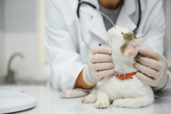
Libby was presented to her primary care veterinarian for a dental prophylaxis. Libby also had a six-month history of panting, polyuria, polydipsia, and recurrent ear infections. Physical examination before the dental procedure revealed a swelling in Libby's perineal region, and the veterinarian subsequently diagnosed a left perineal hernia. The dental prophylaxis was rescheduled for a later date, and Libby was referred to the Mississippi State University College of Veterinary Medicine (MSU-CVM) for hernia repair.
Todd M. Archer, DVM, MS, DACVIM
Referral evaluation
On presentation to MSU-CVM, Libby was bright and alert. The left perineal hernia was noticeable and was easily reduced. Her abdomen appeared distended, but abdominal palpation revealed no abnormalities. The results of a serum chemistry profile revealed mild increases in alanine aminotransferase (136 U/L; reference range = 10 to 90 U/L) and alkaline phosphatase (153 U/L; reference range = 11 to 140 U/L) activities. Libby's urine specific gravity was 1.019. Based on these results and the history of panting, polyuria, polydipsia, and recurrent infections, a workup for hyperadrenocorticism was performed.
Adrenal function test results
Results of an adrenocorticotropic hormone (ACTH) stimulation test revealed a baseline cortisol concentration of 8.5 μg/dl (reference range = 1.4 to 5 μg/dl) and a one-hour post-ACTH cortisol concentration of 24.8 μg/dl (reference range = 5.5 to 20 μg/dl). These results confirmed hyperadrenocorticism.
Imaging
Abdominal imaging was performed to differentiate pituitary-dependent hyperadrenocorticism (PDH) from an adrenal tumor. The abdominal radiographic findings were unremarkable. Abdominal ultrasonography revealed normal-sized and symmetrical adrenal glands and no evidence of an adrenal mass. Also noted were a single hepatic lesion and a single splenic lesion, as well as a gallbladder mucocele in the early stages of development. Ultrasound-guided fine-needle aspirates of the lesions within the liver and spleen were obtained. A thoracic radiographic examination was also performed, and the findings were unremarkable.
Diagnosis
Libby's abnormal ACTH stimulation test results along with the abdominal ultrasonographic findings provided a working diagnosis of PDH.
Cytologic examination of the hepatic and splenic lesion aspirates showed mild extramedullary hematopoiesis, likely consistent with regenerative nodules. The liver aspirates also showed evidence of vacuolar degeneration, a finding consistent with hyperadrenocorticism.
*The dog in the photograph is not Libby, the dog described in this article.
Initial treatment
It was recommended that Libby receive treatment for PDH before undergoing perineal hernia repair surgery, and the owner chose medical therapy for PDH. Management of Libby's mucocele would be instituted later if needed.
Because of Libby's small size and because I start patients with hyperadrenocorticism at the low end of the recommended VETORYL® Capsules (trilostane) dose range, I prescribed 5-mg compounded trilostane capsules, given once daily at a dose of 2.1 mg/kg in an extralabel fashion. These 5-mg capsules were compounded from VETORYL Capsules at the MSU-CVM pharmacy.
Libby's owners were instructed to give Libby one 5-mg trilostane capsule orally once daily in the morning with food and to return in 14 days for a recheck and follow-up ACTH stimulation test. The owners were also instructed to watch for signs of hypoadrenocorticism (such as weakness, lethargy, vomiting, or anorexia) and to seek immediate veterinary care if she exhibited these signs.
Follow-up visits
At the two-week follow-up visit, Libby was doing better at home, with a reduction in clinical signs and no adverse reactions to trilostane. Upon questioning, the owners stated that they had been administering the trilostane capsules at night. The ACTH stimulation test to monitor dogs receiving VETORYL Capsules should be performed four to six hours after drug administration, so the owners were again instructed to administer Libby's trilostane capsules in the morning with food and to reschedule the ACTH stimulation test.
Libby was presented two weeks later for a recheck examination. The owner reported that Libby was doing well at home. Her polydipsia and polyuria had improved slightly, and her panting had decreased markedly. On physical examination, Libby was bright and alert with a normal temperature, pulse, and respiration. Her perineal hernia was unchanged.
An ACTH stimulation test was performed four hours after trilostane administration. Libby's baseline cortisol concentration was 1.9 μg/dl and her post-ACTH cortisol concentration was 3.7 μg/dl, indicating adequate inhibition of glucocorticoid production. Measurement of Libby's serum electrolyte concentrations (recommended at the 30-day post-VETORYL Capsules treatment re-examination) was inadvertently overlooked at this visit.
Libby's owners were instructed to continue to give Libby one 5-mg trilostane capsule once daily in the morning with food, to schedule surgical repair of the perineal hernia within three weeks, and to schedule a three-month post-trilostane treatment recheck examination.
Two weeks later, Libby was presented to MSU-CVM for surgical hernia repair, which was completed without complications. Libby's preanesthetic CBC and serum chemistry profile results were clinically unremarkable. Libby is scheduled to return to the internal medicine service for a three-month post-trilostane treatment reexamination.
Dr. Archer's perspective
Libby's case illustrates the need to evaluate the patient history and results of routine blood tests to help identify hyperadrenocorticism as a possible differential diagnosis. Initially, the dental prophylaxis and perineal hernia were the primary concerns in this patient, but ultimately Cushing's disease was diagnosed and treated.
An early gallbladder mucocele was identified during Libby's workup. The mucocele will be further assessed during future rechecks, with therapy instituted as indicated. In a recent study, mucocele formation was 29 times more likely in dogs with hyperadrenocorticism than in dogs without hyperadrenocorticism.1 Although Libby did not present with clinical signs or have laboratory abnormalities consistent with a gallbladder mucocele, a mucocele should be considered in any dog with hyperadrenocorticism that presents with acute illness and evidence of hepatobiliary disease.
I prescribed compounded trilostane for Libby in an extralabel fashion because of her small size and because I prefer to start patients with hyperadrenocorticism at the low end of the labeled VETORYL Capsules dose range. Specific patients may require trilostane in strengths or forms not offered by VETORYL Capsules. If compounding cannot be avoided, trilostane can only be legally compounded using the contents of FDA-approved VETORYL Capsules as the starting material.2
In a recent study, all batches of capsules compounded from VETORYL Capsules (compounded controls) conformed to the acceptance criteria for trilostane content (90% to 105% of label claim), whereas 38% of the batches of compounded capsules purchased from compounding pharmacies failed to meet the acceptance criteria for content.3 the mean percent dissolution for compounded capsules purchased from compounding pharmacies was also lower and impurity profiles were significantly higher when compared with capsules compounded from VETORYL Capsules. This highlights the need to ensure that the compounding pharmacy you use is technically proficient and formulates compounded trilostane capsules from VETORYL Capsules.
Keep in mind that when the granules are removed from VETORYL Capsules to create a compounded trilostane drug, the drug can no longer be called VETORYL Capsules. Also, the safety, effectiveness, and stability of the compounded drug become the responsibility of the prescribing veterinarian, and are not the responsibility of Dechra Veterinary Products or the compounding pharmacy.4,5
In addition, Libby's case highlights the need to ensure that owners understand how to administer trilostane. The follow-up ACTH stimulation testing that is performed as part of patient monitoring during treatment with VETORYL Capsules should occur four to six hours after administration, which makes administering the drug once a day in the morning ideal.
Dr. Archer is an assistant professor of small animal medicine in the Department of Clinical Sciences at the Mississippi State University College of Veterinary Medicine.
REFERENCES
1. Mesich ML, Mayhew PD, Paek M, et al. Gall bladder mucoceles and their association with endocrinopathies in dogs: a retrospective case-control study. J Small Anim Pract 2009;50:630-635.
2. U.S. Food and Drug Administration. Compliance Policy Guide 7125.40. Section 608.400: Compounding of Drugs for Use in Animals. Available at http://www.fda.gov/ICECI/ComplianceManuals/CompliancePolicyGuidanceManual
/ucm074656.htm. Accessed Sept. 30, 2013.
3. Cook AK, nieuwoudt CD, Longhofer SL. Pharmaceutical evaluation of compounded trilostane products. J Amer Anim Hosp Assoc 2012;48(4):228-233.
4. U.S. Food and Drug Administration. Code of Federal Regulations Title 21. 530.13(b)(4): Rules and Provisions for Extralabel Uses of Drugs in Animals. Available at http://www.accessdata.fda.gov/scripts/cdrh/cfdocs/cfcfr/CFRSearch.cfm?fr=530.13. Accessed Sept. 30, 2013.
5. Spies, A. Compounding: The practice of pharmacy or a liability risk? Drug Topics 2002;14:36. Available at http://drugtopics.modernmedicine.com/drug-topics/news/ce-compounding-practice-pharmacy-or-liability-risk. Accessed Sept. 30, 2013.
This case was solicited from the prescribing veterinarian and may represent an atypical case study. Similar results may not be obtained in every case.
Hyperadrenocorticism affects many adult dogs. Whether the disease is pituitary-dependent (80% to 85% of spontaneous cases) or adrenal-dependent (15% to 20% of cases), the clinical and laboratory abnormalities associated with it result from chronic hypercortisolemia. Clinical signs of hyperadrenocorticism at the time of diagnosis can vary widely, and they develop so gradually that owners often mistake the signs for "normal" aging. Being aware of the more subtle signs of canine hyperadrenocorticism can be key to early diagnosis and initiation of therapy.
Common Clinical Signs of Canine Hyperadrenocorticism
Whenever possible, pituitary-dependent hyperadrenocorticism and adrenal tumors should be differentiated to help guide therapy and patient monitoring. Early diagnosis and management of canine hyperadrenocorticism may not only improve the patient's clinical signs but may also keep the more severe consequences of Cushing's syndrome from developing.
Learn more with these online resources
Go to the Dechra Veterinary Products CE Learning Center at
• Diagnosing and treating canine hyperadrenocorticism
Presented by Audrey K. Cook, BVM&S, MRCVS, DACVIM, DECVIM, and David s. Bruyette, DVM, DACVIM
• Cushing's disease: Inside and out
Rhonda Schulman DVM, DACVIM, and John Angus, DVM, DACVD
• Diagnosing and treating feline hyperthyroidism
Presented by Andrew J. Rosenfeld, DVM, DABVP
Then get your whole team on the same page, by visiting the Team Meeting in A Box section at
• Stop getting burned by ear infections
How you handle otitis externa and ear infections can make or break client bonds—and dogs' well-being. Use this Team meeting in a Box to create a team approach to help pet owners and heal patients.
• Coping with Cushing's syndrome
Pets with Cushing's syndrome suffer from a chronic illness that will be managed throughout the pet's life, not cured. This Team Meeting in A Box will help you deliver a successful team-wide approach.
Visit
Advertisement
Related Content
Advertisement

Latest CME


Advertisement
Advertisement
Trending on dvm360
1
FDA-authorized and approved animal drugs for New World screwworm
2
Hill's introduces therapeutic diets for pets with kidney issues alongside skin or GI sensitivities
3
The human-animal bond was built on parasite control
4
Are you unintentionally sabotaging client visits?
5