

|Articles|November 1, 2009
Based upon the gait, where's the lesion? (Proceedings)
Author(s)Randall C. Longshore, DVM, DACVIM
Observation of gait is an important component of the neurological examination but often is under utilized when assessing patients with gait abnormalities, lameness, or changes in posture.
Advertisement
Observation of gait is an important component of the neurological examination but often is under utilized when assessing patients with gait abnormalities, lameness, or changes in posture. Several factors may interfere with the ability of a veterinarian to properly evaluate the gait of a patient, including space constrictions, flooring that makes gait evaluation difficult, absence of an assistant and time constraints.
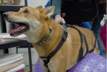
In order to obtain the most information from observing the gait of a patient a space large enough to allow visualization of the patient from the front, back and side is necessary. At a minimum observation of a pet walking in a straight line, turning and walking off a leash is recommended. Watching an animal navigate steps, curbs and other barriers may provide additional information and may be beneficial to further evaluate gait in patients with more subtle deficits. In a busy veterinary practice finding the space to properly evaluate a patient's gait may not be readily available inside the veterinary hospital itself. If necessary a patient can be taken outside to an area of flat, mowed lawn or a non-crowded, cool parking lot or sidewalk. Patient safety and the ability to properly restrain the pet should be kept in mind at all times during the examination both inside and outside the veterinary hospital.
Observation of gait with the patient off a leash is not recommended outside unless a secure, fenced area is available. If observing a pet inside the hospital a room which can be completely secured is also recommended, such as an examination room or large run. Small pets can be adequately observed in even small examination rooms, but for large and giant breed dogs the room should be large enough to allow the patient to move freely and take more than one or two steps. Examination tables that fold against the wall to open up the examination space are useful to allow larger pets a more open area to walk. Ideally the room should be uncluttered and free of low ledges or furniture that a pet can crawl under. This is especially important when observing the gait of feline and exotic animal species.
Animals need secure, comfortable, familiar footing in order to walk and demonstrate their gait most accurately. Many veterinary hospitals have ceramic tile or other hard, smooth surfaces. While these floors make cleaning easy they do not always provide the best footing for patients with weakness or lameness. There are several options to provide more secure footing for patients to facilitate gait observation. Several textured tile and laminate materials are available, as are several types of rubber flooring options. Rubber mats can also be used to temporarily cover areas of less secure footing during examination of gait.
An assistant is recommended to walk the patient while the veterinarian stands back at a comfortable distance. Many pet owners can assist with walking their pet but surprisingly this task is not always as easy as one might think. Ideally the pet will be walked beside the pet owner and not in front or behind them so as to obscure the view of the observer. A short, non-retractable leash should be used to provide the best control of direction and pace of the pet. Keeping short leashes in each examination keeps them readily available and can be used in lieu of the pet owner's retractable leash if necessary. If a pet owner is physically unable or has difficulty controlling their pet the use of a trained veterinary assistant is recommended. This often provides the best, most consistent way to observe the gait of patients.
Finally, in order to obtain the most out of observing a patient's gait the veterinarian has to make the task a priority and make time to evaluate the gait. Often a patient is on the examination table waiting for the veterinarian when he or she enters the examination room. Taking the pet off the exam table, closing the door and removing the leash allows the veterinarian to observe the pet while a history is obtained. Observation of the gait while on a leash should be one of the first components of the examination once a history is obtained if the primary concern is lameness or weakness.
Forebrain (cerebral) syndrome
Patients with disorders of the forebrain may have a normal or near normal gait with good preservation of strength. Patients often have a compulsion to walk (pace), move in a forward direction and have difficulty standing still in one place. As patients pace they may bump into objects or get stuck between chairs, under benches or in the corner of the examination room. Circling may be present in some patients, while others are able to walk in a straight line but turn predominantly in one direction. At times patients may stand with their head turned (not tilted) slightly in the direction in which they turn or circle. Confusion or a blank/distant expression is also present in some patients. When walking in a straight line on a leash patients with forebrain disease may tend to drift to one direction. Drifting to one side can also be seen in patients with vestibular or brainstem disease, but animals with forebrain disease are typically stronger and less ataxic than patients with vestibular or brainstem symptoms. As forebrain symptoms continue to advance in severity patients do become weaker, but other symptoms of forebrain disease such as confusion, depression and dull behavior are also more evident.
Brainstem syndrome
The brainstem is an extension of the spinal cord and as such disease of the brainstem causes much more severe weakness than forebrain disease. Tetraparesis (often asymmetric) or hemiparesis ipsilateral to the side of the lesion is present and patients may knuckle on one side of their body. Some patients with brainstem disease will have a head tilt, visible nystagmus and a tendency to lean or fall to one side if the central vestibular system is involved. Behavior changes or altered mentation may also occur in patients with an acute onset or severe brainstem disease.
Advertisement
Cerebellar syndrome
The classic gait of a patient with cerebellar disease is dysmetria (such as hypermetria of the front limbs), spastic, exaggerated limb movements and a wide based stance when not moving. Some patients also sway and have truncal ataxia when trying to maintain a still posture. Intention tremors of the head are often noticeable when patients sniff objects or attempt to pick items up in their mouth. Patients with pure cerebellar symptoms have preserved strength, do not knuckle as they walk and have normal or near normal mentation. Often, however, patients with symptoms of cerebellar disease also have some degree of brainstem compression. In these patients hemiparesis or asymmetric tetraparesis and altered mentation may also be present.
Cervical syndrome (C1-C5)
Patients with spinal cord disorders from C1-C5 have gaits that range from normal to hemiparesis/plegia or non-ambulatory tetraparesis/plegia. If pain is present most patients will guard their neck and limit voluntary movement of the head in one or all directions. The head is lowered and the neck either extended or retracted. In short haired breeds, visible twitching or fasciculation of the cervical muscles may be present, especially when animals move their head and neck. As symptoms progress patients knuckle in the front and/or rear limbs. Despite these symptoms, mentation of patients with cervical symptoms is normal.
Cervicothoracic syndrome (Cervical Enlargement C6-T2)
Weakness in animals with C6-T2 spinal cord disease may occur in all limbs, one side of the body or in a single limb (monoparesis). Disorders of this area of the spinal cord also may produce the classic "two engine" gait in which the front limbs move in quick, spastic movements while the rear limb gait is longer and characterized by marked ataxia. In yet other patients with C6-T2 spinal cord disease the front limbs may be weaker than the rear limbs. Neck guarding and muscle fasciculations may also occur, as well as muscle atrophy to the front limbs. When nerve root compression is present in this area many patients will flex a front limb to the body. This posture is called the root signature sign.
Thoracolumbar syndrome (T3-L3)
Patients with spinal cord disease from T3-L3 have normal strength and gait in the front limbs but varying degrees of rear limb weakness. Some patients have normal rear limb strength but have back pain and often arch their back. Increased extensor tone is often present and may produce a rigid rear limb gait in patients that are still ambulatory. Non-ambulatory patients may sit directly on their perineal area and have the rear limbs extended stiffly in front of their body.
Lumbosacral syndrome and cauda equina (Lumbar Enlargement L4-Cd5)
Symptoms produced by lesions from L4-Cd5 vary depending on the location within the lumbar enlargement and the size of the patient. In large breed dogs the filum terminale of the spinal cord may be as far cranial as L5. In small breed dogs and cats, however, the filum terminale extends into the sacrum. Therefore, some patients will experience only pain while others will have rear limb weakness or paralysis. Muscle tone to the rear limbs may be diminished, giving paretic animals a crouched stance or gait. Decreased tail movement or paralysis may be observed. This is most easily recognized in cats or dogs that normally carry the tail elevated or over their back. In other patients the tail may swing from side to side in a pendular motion as they walk. A rear limb root signature sign may be present and is especially common with lesions at L5-L6 and L6-L7. Urinary and fecal incontinence may also be observed as a patient walks on some occasions. Front limb strength and mentation are normal.
Vestibular syndrome
Patients with vestibular disease have a head tilt and may lean or drift to one side as they walk. Others walk in circles or roll if lifted into a standing position. Many patients with vestibular disease become tense and exhibit increased extensor tone when they are lifted off the ground and they loose the ability to compensate for their dizziness with the sense of touch. When standing patients with vestibular symptoms may lean or have a wide based stance. With peripheral vestibular disease mentation is normal, however, with central vestibular disease altered mentation may be present.
Neuromuscular syndrome
Neuromuscular disorders produce weakness that is made worse with movement and exercise. After rest patients with neuromuscular disease may not have any gait deficits but as they continue to walk their length of stride shortens giving the appearance of a short, choppy gait. If forced to continue walking weakness worsens and patients may appear lame as they try to rapidly take weight off a limb, not because of pain but because of weakness. Patients may drop their head as the cervical muscles weaken and if they continue may "nose dive" and eventually collapse in the front limbs. Many patients with neuromuscular weakness are reluctant to stand still and prefer to either walk or lie down. When standing muscle tremors in the limbs may be present. Despite sometimes marked weakness, most patients with neuromuscular symptoms have little to no ataxia. Increased inspiratory stridor when panting or excited may be evidence of laryngeal paralysis and neuromuscular weakness.
Orthopedic disorders
It is sometimes very difficult to distinguish between lameness produced by certain orthopedic disorders and neurologic conditions. Gait abnormalities with orthopedic disorders are often worse after a patient has been immobile for a period of time and may improve substantially after a few steps. Orthopedic disease is also more likely to cause a patient to carry the limb for multiple steps or while standing. Proprioceptive placing should also be normal in patients with orthopedic injuries so if a patient is observed knuckling on a limb as they walk a neurologic disorder should be suspected.
Some of the most common disorders that are especially challenging to distinguish between orthopedic and neurologic disorders are peripheral nerve (e.g. brachial plexus) tumors, bilateral ruptured cranial cruciate ligaments and polyarthritis. Patients with polyarthritis and bilateral ruptured cranial cruciate ligaments may be unable to walk or walk with a very stiff, unsteady gait. With close observation one can see that the limbs are picked up off the ground or slid along the ground with each step, rather than having the toes drag. Proprioceptive placing is normal in patients with polyarthritis and bilateral ruptured cranial cruciate ligament rupture. Lameness produced by peripheral nerve tumors may be especially difficult to isolate, especially early in the course of the disorder. Peripheral nerve sheath tumors should be suspected in patients with progressive lameness in which no other cause can be identified.
Acknowledgement
The title of this lecture was inspired by my first neurology mentor Dr. Robert Selcer. I am forever grateful to Dr. Selcer for his ability to make the complexities of neurology understandable and less intimidating for me and the hundreds of veterinary students fortunate enough to have learned neurology through his eyes. I am especially grateful for the encouragement he provided at a very formative part of my training.
Advertisement
Related Content
Advertisement


Latest CME


Advertisement
Advertisement
Trending on dvm360
1
Q&A: Managing anemia, appetite loss, and phosphorus in feline chronic kidney disease
2
Weekly Vet Report: FIP's shorter treatment window, lone star ticks driving red meat allergies & more
3
Pet ownership and care should not only be for the wealthy
4
What the new AAHA Diabetes Management Guidelines for Cats say about SGLT2 inhibitors
5