

|Articles|August 1, 2010
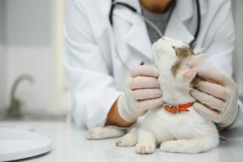
Approach to jaundice (Proceedings)
Jaundice (or icterus), broadly defined, refers to the accumulation of excessive amounts of bilirubin in either the circulation or the tissues.
Advertisement
Jaundice: Jaundice (or icterus), broadly defined, refers to the accumulation of excessive amounts of bilirubin in either the circulation or the tissues.
Normal Bilirubin Metabolism (see Figure 1)
Bilirubin is primarily derived from breakdown of the hemoglobin of aging red cells by the mononuclear phagocytic system (MPS). Although the normal degradation of erythrocytes can occur in any tissue that contains phagocytes, the process is concentrated in the organs of the MPS (particularly the spleen). The hemoglobin molecule is initially split into heme and globin. Heme is further metabolized into biliverdin and eventually bilirubin, during which process the iron within the heme molecule is extracted for either reuse in the production of hemoglobin or storage within tissues. Smaller amounts of bilirubin are obtained from the degradation of other iron-containing molecules such as myoglobin and hepatic hemoproteins. Bilirubin is released from the tissues into the blood stream, where it binds to albumin and is carried via the circulation to the liver. Albumin-bound bilirubin (called unconjugated, indirect or free bilirubin) is fat soluble and water insoluble, and cannot pass from the renal circulation into the urine. Unconjugated bilirubin, upon reaching the hepatic vasculature, is separated from albumin and enters hepatocytes. The hepatic enzyme glucuronyl transferase conjugates intracellular unconjugated bilirubin to form bilirubin diglucuronide (called conjugated or direct bilirubin). Conjugated bilirubin is then released from hepatocytes into the bile canaliculi, and excreted via the biliary system into the duodenum. Hepatocyte release of conjugated bilirubin into bile is the rate-limiting step of bilirubin metabolism. Hepatocytes do not normally release significant amounts of conjugated bilirubin back into the circulation. Conjugated bilirubin is water soluble and fat insoluble, and when present in the circulation will enter the urine. Conjugated bilirubin entering the ileum and large intestine from the duodenum is converted by bacterial enzymes into urobilinogen, most of which is subsequently excreted the feces. Bile-derived pigments such as urobilin (oxidized urobilinogen) give feces its characteristic color. Some intestinal urobilinogen is reabsorbed by the colon and enters the portal circulation. Although most of this reabsorbed urobilinogen is recycled by the liver, small amounts enter the systemic circulation and spill over into the urine.
Figure 1: Normal Bilirub in Metabolism
Classification of Jaundice
Jaundice is classified as prehepatic, hepatic or posthepatic, a classification based on the phase in bilirubin metabolism that is affected by the causative disease process.
1. Prehepatic
Prehepatic jaundice is usually caused by the release of hemoglobin associated with RBC lysis. High plasma levels of unconjugated (free) bilirubin derived from massive hemoglobin breakdown can overwhelm hepatic bilirubin metabolism. Since the liver is usually very effective at conjugating free bilirubin, hemolysis must be acute and severe to cause jaundice. Patients with prehepatic jaundice are therefore invariably severely anemic. Jaundice is typically transient, since ongoing hemolysis of sufficient magnitude to cause persistent jaundice would cause death due to anemia. Hemolytic anemia in dogs and cats is most commonly subacute or chronic, and therefore usually does not cause detectable jaundice.
Theoretically, prehepatic jaundice should cause high unconjugated bilirubin and normal conjugated bilirubin levels. However, the increased hepatic production of conjugated bilirubin in response to high levels of unconjugated bilirubin can overwhelm hepatocyte excretion mechanisms, particular if liver function is impaired by hypoxia secondary to severe anemia. Prehepatic jaundice is therefore often a mixture of unconjugated and conjugated hyperbilirubinemia.
Common Causes of Prehepatic Jaundice
Immune-mediated hemolytic anemia Zinc toxicity
Heinz-body hemolytic anemia (e.g. onion toxicity) Erythrocyte parasites
Transfusion reaction
Cats: Mycoplasma hemofelis infections Dogs: Heartworm (caval syndrome)
Feline leukemia virus-associated anemia
Hypophosphatemia-induced hemolytic anemia
Acetaminophen toxicity
Neonatal isoerythrolysis
2. Hepatic Jaundice
Hepatic jaundice is caused by dysfunction of either hepatocytes or the intrahepatic biliary tree. Hepatocyte uptake and conjugation of free bilirubin is impaired, as is hepatocyte and biliary excretion of conjugated bilirubin. Since the healthy liver has a large functional reserve, most normal hepatic functions (including bilirubin metabolism) are not impaired unless liver disease is severe. Mild hepatic diseases such as steroid or diabetic hepatopathy do not typically cause jaundice. Theoretically, any cause of severe liver dysfunction could cause hepatic jaundice. However, despite causing profound hepatic dysfunction, some diseases (portosystemic shunts in particular) rarely cause jaundice. Since hepatocyte excretion of conjugated bilirubin is the rate-limiting step in bilirubin metabolism, hepatic jaundice would be expected to be associated with elevated plasma levels of conjugated bilirubin and normal unconjugated bilirubin levels. Usually, however, the intracellular build up of conjugated bilirubin in the failing liver then causes unconjugated bilirubin to also bank up within hepatocytes, following which both forms of bilirubin spill over into the circulation. Like prehepatic jaundice, hepatic jaundice is therefore usually a mix of unconjugated and conjugated hyperbilirubinemia.
Common Causes of Hepatic Jaundice
Toxic hepatopathy Hepatic neoplasia (primary or metastatic)
Hepatic amyloidosis
Cats: Suppurative or lymphocytic cholangiohepatitis Dogs: Chronic hepatitis
Idiopathic hepatic lipidosis Copper storage diseases
FeLV-associated diseases (esp. lymphosarcoma) Leptospirosis
Feline infectious peritonitis Infectious canine hepatitis
Toxoplasmosis Hepatic fibrosis/cirrhosis
Acetaminophen toxicity Thiacetarsamide
3. Posthepatic Jaundice
Posthepatic jaundice is caused by either obstruction or rupture of the biliary tree, with impairment of excretion of conjugated bilirubin into the small intestine. Since bilirubin pigments such as urobilin cannot enter the intestine if obstruction or rupture is complete, feces may become gray ('acholic' feces). Biliary obstruction or rupture can cause severe jaundice in the presence of normal hepatocellular function. Posthepatic jaundice typically develops gradually, most often over a period of days or even weeks. Jaundice secondary to biliary obstruction is initially associated with elevated plasma levels of conjugated bilirubin and normal unconjugated bilirubin levels. However, since the liver cannot excrete excess bilirubin via the biliary tree, conjugated and then unconjugated bilirubin bank up within hepatocytes and spill into the circulation. Conjugated bilirubin can also spontaneously deconjugate within the circulation. Chronic cases of biliary obstruction may therefore present with a mixture of unconjugated and conjugated hyperbilirubinemia. Chronic obstruction will also eventually directly impair hepatocellular function (probably due to the build up of hepatotoxic bile acids), leading to a combination of hepatic and posthepatic jaundice. Rupture of the biliary tree with resultant bile peritonitis will cause progressing conjugated hyperbilirubinemia as bilirubin diffuses into the circulation from the peritoneal cavity.
Common Causes of Posthepatic Jaundice
Biliary Obstruction: Pancreatitis (acute or chronic) with compression of the bile duct
Neoplasia (intestinal, pancreatic or hepatic) with bile duct compression
Cholelithiasis or inspissated bile
Biliary Rupture: Traumatic rupture of bile duct or gall bladder
Clinical Signs Associated With Jaundice
Tissue jaundice that is obvious on physical examination is usually associated with marked hyperbilirubinemia (total serum bilirubin of at least 2 mg/dl). Hyperbilirubinemia is therefore detectable as a biochemical abnormality long before tissue jaundice becomes apparent. Serum jaundice will usually also be obvious on examination of the supernatant of a centrifuged blood sample well before overt tissue jaundice develops. Since conjugated bilirubin is water soluble and diffuses into tissues much more readily than unconjugated bilirubin, hepatic or posthepatic jaundice (in which conjugated hyperbilirubinemia initially predominates) may be detectable as tissue jaundice at lower total serum bilirubin levels than prehepatic jaundice.
Subtle jaundice may be detected by close inspection of the sclera, the inside of the pinnae, the back of the throat and the skin of the ventral abdomen. Since the magnitude of tissue jaundice usually reflects the degree of hyperbilirubinemia, serum bilirubin levels are rarely normal in the presence of obvious tissue jaundice except on those uncommon occasions when tissue levels of bilirubin remain elevated for a few days after bilirubinemia has resolved. Since the renal threshold for bilirubin is low, particularly in dogs, most jaundiced patients have marked bilirubinuria and orange urine. Theoretically, patients with acute hemolysis and early prehepatic jaundice (elevated free bilirubin and normal conjugated bilirubin levels) may not have bilirubinuria since free bilirubin is bound avidly to albumin and cannot enter the urine.
Jaundice is usually well-tolerated, and patients presenting with isolated jaundice (for example, those with biliary obstruction or rupture and normal hepatocellular function) are often remarkably unaffected. Profound elevations in unconjugated bilirubin (observed, for example, in neonatal isoerythrolysis, a rare entity in dogs and cats) can cause neurotoxicity and nephrotoxicity ('kernicterus'). The signs observed in jaundiced patients, however, usually reflect the consequences of underlying or concurrent diseases (such as hemolytic anemia or liver failure) rather than the effects of the jaundice itself.
Prehepatic Jaundice
Patients with obvious prehepatic jaundice usually have severe hemolysis, and the clinical signs of anemia (weakness, pale mucous membranes, tachycardia and tachypnea) tend to predominate. Severe intravascular hemolysis may cause hemoglobinemia and hemoglobinuria ('port wine' urine).
Hepatic Jaundice
Since patients with jaundice due to hepatic disease invariably have profound hepatic dysfunction, jaundice usually occurs concurrently with other signs associated with liver failure such as weight loss, anorexia, ascites, vomiting, diarrhea, polydipsia/polyuria, coagulopathy and hepatoencephalopathy.
Posthepatic Jaundice
Patients with biliary obstruction often remarkably unaffected for long periods (days to weeks). The presence of pale gray (acholic) feces suggests that obstruction is complete. Since bile acids excreted into the duodenum via the biliary tree are needed for fat absorption, biliary obstruction can cause fat malabsorption and steatorrhea (greasy feces). Fat malabsorption may cause decreased absorption of fat-soluble vitamins, particularly vitamin K. Patients with chronic biliary obstruction are therefore prone to bleeding disorders due to depletion of the active forms of the vitamin K dependent clotting factors (II, VI, IX and X). Patients with biliary obstruction due to acute pancreatitis may exhibit anorexia, vomiting, diarrhea and abdominal pain. Obstruction secondary to chronic pancreatitis with pancreatic scarring or abscessation, in contrast, may occur months or even years after the last clinically apparent episode of typical pancreatitis. In fact, in some patients, pancreatitis remains entirely subclinical, and jaundice is the only presenting complaint. Patients with biliary obstruction secondary to neoplasia may exhibit signs consistent with cancer, such as anorexia or weight loss.
Since the jaundice associated with biliary rupture occurs following the resorption of bile pigments accumulating in the peritoneal cavity, patients with this form of posthepatic jaundice invariably have marked ascites. Bile peritonitis is often well tolerated, and may be associated with minimal malaise, anorexia, pyrexia or abdominal pain unless the peritoneal fluid is becomes infected.
Diagnosis
The serum biochemistry, urine analysis and gross inspection of feces.standard diagnostic testing indicated in patients presenting with jaundice usually includes hematology,
Hematology
Hematology alone is often enough to diagnose prehepatic jaundice, since hemolysis must invariably be severe before jaundice becomes apparent (PCV of less than 15-20% in the dog, and 10-15% in the cat). Since less severe anemia is unlikely to cause clinically detectable jaundice, mild to moderate anemia in a jaundiced animal is most likely to be due to the disease process causing the jaundice (for example, the anemia of chronic disease).
Serum Biochemistry
Biochemistry may suggest ongoing hepatic damage (elevated ALT or ALP) or dysfunction (hypoalbuminemia, hypoglycemia and decreased urea). Biochemical tests that may need to be specifically requested, since they are not always included in the profiles provided by laboratories, include amylase, lipase and total serum bilirubin.
Urinalysis
Since dogs have a low renal threshold for conjugated bilirubin, it is common to detect small amounts of bilirubin and sporadic bile crystals in normal, concentrated canine urine. The detection of higher levels of urinary bilirubin (2+ or more on dipstick), particularly in dilute urine, suggests increased serum levels of conjugated bilirubin (hepatic or posthepatic jaundice). Since the canine renal tubules can conjugate some bilirubin, bilirubinuria may also potentially occur with prehepatic jaundice even though protein-bound unconjugated bilirubin cannot pass directly into the urine. Cats, in contrast to dogs, have a high renal threshold for conjugated bilirubin. Detection of any amount of bilirubin in cat urine is therefore always clinically significant, and should be investigated.
Feces
The presence of acholic feces or steatorrhea is highly suggestive of posthepatic jaundice due to complete biliary obstruction. Partial biliary obstruction, however, is usually associated with normal fecal color and composition.
Diagnostic Features of Prehepatic, Hepatic and Posthepatic Jaundice
After evaluation of hematology, biochemistry, urine and feces, the ensuing approach to jaundice is facilitated by an understanding of the diagnostic features of prehepatic, hepatic and posthepatic jaundice.
1. Prehepatic Jaundice
Prehepatic jaundice is invariably associated with severe hemolysis, and intravascular hemolysis may also cause hemoglobinemia and hemoglobinuria. In the dog, prehepatic jaundice is usually due to severe acute forms of immune-mediated hemolytic anemia, whereas in the cat the more common causes of prehepatic jaundice also include Mycoplasma hemofelis and hemolysis associated with FeLV infection. Specific tests commonly indicated in the small animal patient with prehepatic jaundice therefore include:
Visual inspection of serum and urine color
Rapid slide agglutination and/or Coomb's test
Examination of red cells for Mycoplasma hemofelis organisms (cats)
Serological testing for FeLV (cats)
Less common forms of hemolysis, such as Heinz-body hemolytic anemia, acetaminophen toxicity, zinc toxicity, hypophosphatemia-induced hemolysis, transfusion reactions, neonatal isoerythrolysis and caval syndrome can usually be determined by history, routine diagnostic testing and more specific tests such as serum phosphate, heartworm testing, and abdominal radiography.
2. Hepatic Jaundice
Over half of the original functional hepatocellular mass must be lost before dysfunction is detectable using standard function tests. Dysfunction must be even more advanced to produce the classic signs and biochemical derangements (jaundice, hypoalbuminemia, hypoglycemia and low urea) associated with hepatic failure. By the time a patient with hepatic disease becomes jaundiced, sensitive hepatic function tests such as ammonia tolerance will almost invariably be abnormal. Standard function tests which may be available include BSP excretion, oral or rectal ammonia tolerance testing, and pre- and post-prandial serum bile acids. Of these tests, only ammonia tolerance is likely to be of diagnostic value in patients with jaundice, since ammonia metabolism is a hepatic function that is not dependent on biliary excretory pathways. BSP excretion and bile acids are usually abnormal in animals with hepatic or posthepatic jaundice, since both tests are dependent on biliary excretory mechanisms. BSP excretion and bile acids therefore provide little extra diagnostic information in the jaundiced patient.
Liver enzymes such as ALT, ALP and GGT are often elevated in patients with hepatic jaundice. Since ALT is a 'leakage' enzyme that is liver-specific, elevated ALT indicates active hepatocellular damage. ALP and GGT, in contrast, are 'induced' enzymes, and elevations in these enzymes can occur without ongoing cell damage. Both ALP and GGT are induced by cholestasis, and will be elevated with either extrahepatic (bile duct obstruction) or intrahepatic (diffuse obstruction of bile canaliculi) cholestasis. Since primary hepatic diseases (such as lipidosis) can cause diffuse obstruction of canaliculi secondary to hepatocyte swelling, elevations in ALP or GGT do not always indicate that the disease is centred on the biliary tree. ALP is not liver-specific (particularly in dogs), as various non-cholestatic isoenzymes can also be induced by steroids, anticonvulsants and bone growth. Compared to the dog, liver enzymes have much shorter circulating half lives in the cat. Any elevation in ALT or ALP in the cat should therefore be regarded as diagnostically significant. ALP and GGT are usually concurrently elevated in cats with hepatic disease. Paradoxically, lipidosis often causes marked elevations in ALP despite normal GGT. Theoretically, elevations in liver enzymes (particularly ALT) should distinguish hepatic from prehepatic and posthepatic jaundice. However, since both hypoxia (due to severe anemia) and accumulating serum bile acids (due to obstruction) can cause hepatocellular damage, enzymes can be elevated in all three forms of jaundice. Furthermore, patients with hepatic jaundice due to severe chronic liver disease may have so few hepatocytes remaining that serum levels of liver enzymes will return to normal despite ongoing hepatic damage.
Since the liver synthesizes most of the clotting factors, coagulopathies are common in patients with hepatic failure. Assessment of the intrinsic and extrinsic coagulation pathways via measurement of PTT and PT respectively is often indicated in patients with hepatic jaundice, particularly prior to invasive procedures such as liver biopsy. Since chronic biliary obstruction can cause depletion of the active forms of the vitamin K dependent factors secondary to malabsorption of fat-soluble vitamins, PT and PTT may also be prolonged in patients with posthepatic jaundice. Correction of PT and PTT after supplementation with parenteral vitamin K suggests that the coagulopathy is due to biliary obstruction rather than hepatocellular failure. Liver size/architecture may be evaluated by abdominal radiography and ultrasonography. Depending on the cause of hepatic failure, the liver may be small (chronic hepatitis or cirrhosis), large (neoplasia, amyloidosis or lipidosis) or of normal size. Microhepatia (small liver size) is rarely observed in cats with liver failure.
3. Posthepatic Jaundice
Posthepatic jaundice due to extrahepatic biliary obstruction is notoriously difficult to distinguish from hepatic jaundice secondary to liver failure, particularly as both syndromes commonly cause elevated liver enzymes. Since obstruction is often associated with a better prognosis than hepatic failure, it is important to be able to confidently differentiate the two conditions if possible. Diagnostic tools that may be useful include:
Serum Biochemistry
Elevated amylase and lipase are most consistent with obstruction secondary to pancreatic disease, whereas hypoalbuminemia, low urea and hypoglycemia are more suggestive of hepatic failure.
Liver Function Tests
Ammonia tolerance is usually abnormal in patients with hepatic jaundice. Patients with posthepatic jaundice, in contrast, often have normal ammonia tolerance, particularly early in the disease process. Chronic obstruction, however, may lead to hepatic dysfunction due to the toxic effects of bile acids.
Response to Vitamin K
Correction of PT or PTT with parenteral vitamin K is suggestive of complete biliary obstruction.
Abdominal Radiography and Ultrasonography
Detection of microhepatia via radiography or ultrasonography strongly suggests the presence of chronic liver failure. Ultrasonography may also be able to differentiate parenchymal disease from biliary obstruction, and may even reveal the cause of obstruction (pancreatic disease, cholelithiasis).
Laparotomy/Laparoscopy
Liver biopsy is sometimes needed to differentiate hepatic from posthepatic jaundice. Laparotomy is often the best means of obtaining a biopsy, since laparotomy also enables assessment of the patency of the bile ducts and immediate surgical correction of biliary obstruction if necessary. Recently, some referral centers have begun offering laparoscopy as an alternative means of visualizing and biopsying the liver.
Posthepatic jaundice secondary to biliary rupture is usually easy to identify. Affected patients typically have ascites, and often have a history of abdominal trauma. Suspected biliary rupture and bile peritonitis can be confirmed by abdominocentesis. Aspirated fluid usually has a characteristic dark green color.
Liver Biopsy
Cytologic or histopathologic examination of hepatic tissue is essential for establishing a definitive diagnosis in most cases of primary hepatic disease. Options available for obtaining tissue samples include:
• Fine needle aspiration cytology
• Blind or keyhole percutaneous needle biopsy
• Ultrasound-guided percutaneous needle biopsy
• Laparoscopic biopsy
• Exploratory laparotomy and wedge biopsy
Hepatic biopsy via laparotomy is indicated in patients with focal liver disease, microhepatia, ascites or coagulopathies, or when either bacterial infection (such as suppurative cholangiohepatitis) or biliary obstruction is considered to be possible diagnoses. In such patients, percutaneous aspiration or biopsy techniques risk obtaining non-diagnostic samples or causing iatrogenic hemorrhage, bacterial peritonitis or bile leakage. Percutaneous biopsy techniques, however, avoid unnecessary invasive surgery, and are relatively safe in patients with accessible livers (normal or enlarged in size), adequate hemostasis and diffuse hepatic parenchymal disease. Percutaneous biopsy techniques may be safely performed with heavy sedation and local anesthesia in tractable patients. Percutaneous biopsies, however, should not be performed if the clinician feels that subsequent laparotomy is likely to be needed either to collect further samples (bile for bacterial culture, for example) or to institute therapy (for example, bile duct repair or gastrostomy tube placement in cats with hepatic lipidosis).
Advertisement
Related Content
Advertisement

Latest CME


Advertisement
Advertisement