

|Articles|August 1, 2009
Urolithiasis in small ruminants (Proceedings)
Obstructive urolithiasis is considered to be the most economically significant urinary tract disease of food animals, affecting primarily intact and castrated male ruminants, swine and camelids.
Advertisement
Obstructive urolithiasis is considered to be the most economically significant urinary tract disease of food animals, affecting primarily intact and castrated male ruminants, swine and camelids. Male small ruminants are particularly predisposed, while females are rarely clinically affected.
Pathogenesis
Uroliths are solid crystalline formations in the urine which are composed of organic matrix and organic and inorganic crystalloids. Matrix, made up of sugars, proteins, and cells, results from urine super-saturation. Factors affecting urine super-saturation include the rate of renal excretion of crystalloids, negative water balance, urine pH and the presence or absence of crystallization inhibitors. Metaplasia of uroepithelium, as a result of vitamin A deficiency, may contribute cells and protein for nuclear formation. Suture, tissue debris, blood clots or bacteria may also serve as nuclear components initiating urolith formation. Infection, however, is considered to be a minor factor in urolith formation in ruminants. The formation of a nucleus is followed by deposition of inorganic minerals, including magnesium, calcium and phosphate, onto the matrix.
The anatomy of the distal urinary tract of male ruminants differs significantly from that of males of other species and contributes to the development of obstruction and increases treatment difficulty. The penis is sigmoid in arrangement, with two major bends occurring between the urinary bladder and the distal glans penis. The distal flexure is a common site of urethral obstruction by uroliths. The glans penis of the small ruminant also has a vermiform appendage, or urethral process, which is an extension of the urethra 2-4 cm beyond the distal end of the penis. It has a narrowed diameter compared to the more proximal portions of the urethra and also serves as a common location for obstruction. The urethral diverticulum, an outpouching of the urethra at the level of the ischial arch, complicates treatment of affected animals. When a urinary catheter is passed into the urethra in a retrograde manner from the glans in order to access the urinary bladder, this diverticulum readily accepts the catheter, rather than allowing the catheter to proceed into the urinary bladder.
Urolithiasis is a multifactorial disease with such inputs as diet, urine pH and body water balance. Struvite (magnesium ammonium phosphate) and apatite (calcium phosphate) may be commonly seen in animals fed high-grain diets, while animals consuming legumes are predisposed to calcium carbonate uroliths. Silicate stones may be observed in animals grazing silicaceous plants and soils in the western United States and Canada. Calcium oxalate stones may be associated with oxalate containing plants.
A significant factor in availability of urolith components and their binding ability is urine pH. Struvite, apatite and calcium carbonate uroliths are known to precipitate in alkaline urine. Struvite crystallization occurs only at a pH range of 7.2 – 8.4, while apatite stones develop at a urine pH of 6.5 – 7.5. Calcium carbonate stones also tend to form in alkaline urine, while pH may have little or no effect on silicate or calcium oxalate uroliths.
Total body water balance plays an important role in calculogenesis by its effects on urine volume and concentration. This may be seen in winter and during times of other systemic illness, when animals consume decreased volumes of water. A negative body water balance contributes to super-saturation, precipitation and formation of residue of organic and inorganic crystalloids in urine.
Uroliths may obstruct urine flow anywhere from the renal pelvis to the distal urethra, although the most common sites of obstruction are at the distal sigmoid flexure or the vermiform appendage in sheep and goats. Obstruction at these sites may result in either rupture of the urethra or of the urinary bladder.
Treatment
Management of affected individuals consists of establishing a patent route of urine excretion, providing analgesia, correcting fluid deficits and correcting electrolyte derangements, decreasing inflammation and preventing infection.
The presence of the urethral diverticulum prevents passage of a urinary catheter retrograde from the urethral orifice to the urinary bladder. Retrograde catheterization or retropulsion of uroliths is not recommended to avoid further trauma or puncture of the urethra at the level of the diverticulum. Attempts at retropulsion of uroliths may result in over distention of the urinary bladder as the stone is diverted into the diverticulum, allowing fluid to pass into the bladder, followed by the urolith falling back into the urethra and preventing the bladder from emptying. Occasionally removal of the vermiform appendage in small ruminants establishes a patent urethra; however inflammation in the proximal urethra from passage of the uroliths may still prevent normal urination. Uroliths tend to occur in multitudes in the urinary bladder and 80% of animals initially relieved by amputation of the vermiform appendage will reobstruct with subsequent stone passage. Relief of urinary obstruction most often requires surgical intervention.
Sedatives may be useful to facilitate treatment. Historically, acepromazine (0.05-0.1 mg/kg, IV or IM) has been utilized in the medical management of urolithiasis. Unproven arguments for utilization of acepromazine have been to relax urethral tone through a-antagonistic effects on smooth muscle and relaxation of the retractor penis muscle. Benefits of acepromazine may also include suppressing the anxiety associated with the inability to urinate. Caution should be taken when using phenothiazine tranquilizers in patients which may already be hypotensive and hypothermic. Diazepam (0.1 mg/kg, slow IV) may also be used for urethral relaxation and as an anxietolytic. Xylazine (0.05-0.1 mg/kg, IV or IM) or other a-2 agonists may be used in attempt to restrain the patient for examination of the penis and have excellent analgesic properties in ruminants. Caution should be exercised when utilizing xylazine prior to relief of the obstruction, as it promotes diuresis, as well as enhancing hypotension. Lumbosacral epidurals using 2% lidocaine (1mL/7kg) may be utilized in the place of sedation to relieve discomfort and aid in exteriorization of the penis.
Fluid therapy should be instituted as indicated by the clinical findings and economics of the case. After relief of the obstruction, diuresis is important to replace dehydration, reduce azotemia and flush the urinary tract. 0.9% NaCl is a good choice for intravenous fluid therapy, although additional electrolyte and acid-base abnormalities should be considered.
Non steroidal anti-inflammatory drugs should be administered to decrease inflammation and aid in the prevention of urethral stricture formation, but should be used with caution until adequate renal perfusion is attained. Broad-spectrum antibiotic therapy should be instituted to prevent or treat infection resulting from devitalized or inflamed urinary tissues or cavitational accumulation of urine. Beta-lactams (penicillins and cephalosporins) may be chosen, as they have good spectrum of activity and are excreted in the urine.
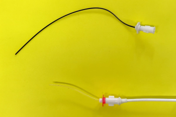
There are many surgical options available, usually selected based upon economic constraints and purposes of the animal. Animals with ruptured urethras should be considered lost for breeding purposes due to adhesion formation. Salvage procedures include perineal urethrostomy and penile amputation, both of which typically suffer from stricture formation weeks to months after surgery. Tube cystotomy is the most successful method of urine diversion, where a foley catheter is placed in the urinary bladder, passes through the abdominal wall and drains externally. This diversion allows for urethral healing and stone dissolution through flushing or diet change and tubes are left in place until urine flow is achieved through the urethra. Other options include urinary bladder marsupialization, urethrotomy and laser lithotripsy. Each of these methods is described in most food animal surgery texts.
Advertisement
Once the obstruction is relieved, treatments to acidify the urine should be initiated in an effort to solubilize additional stones and sediment. Ammonium chloride at a dosage of 200 mg/kg may be orally administered initially and adjusted to attain a urine pH of 6.0-6.5. Care should be taken in dosing so that systemic over-acidification does not occur.
Prevention
Due to the poor prognosis and expense associated with clinical cases of obstructive urolithiasis as well as the herd or flock implications of the disease, considerable focus should be placed on prevention. The important role of metabolic by-products and minerals in the pathophysiology causes diet to be the primary focus of disease prevention. Risk factors addressed in preventative strategies include high dietary phosphorus relative to calcium, high magnesium, low fiber content of rations, low urine output and an alkaline urine pH. Additional factors including selective grazing and castration timing may be addressed.
An elevated level of phosphorus in the diet, with a calcium:phosphorus ratio less than 2:1 increases the excretion of phosphorus in the urine and provides an ion to bind to organic matrix. Increasing the level of calcium in the diet markedly decreases the incidence of urolithiasis, probably due to competition with phosphorus for intestinal absorption and matrix binding. Phosphorus should not comprise greater than 0.6% of the total ration8 and it is recommended that a 2.5:1 or 2:1 calcium:phosphorus ratio be achieved, by the use of calcium salts, if necessary. Calcium oversupplementation should be avoided as increased calcium excretion in the urine may contribute to calcium-containing uroliths.1 High phosphorus levels are present in grains, particularly sorghum, wheat, corn, milo and oats.
A reduction in phosphorus excretion into the urine is also desirable. Ruminants excrete phosphorus primarily by saliva, where it is then swallowed and removed from the body in the feces. Excessive dietary levels of phosphorus may saturate this salivary pathway, causing the excess to be excreted in the urine. Urine phosphorus excretion is greater in animals fed pelleted rations as compared to meal-type rations, due to a decrease in saliva production, and therefore a pathway for excess phosphorus excretion. Increases in the roughage component of diets are important from this standpoint as they increase the amount of saliva that must be produced for proper mastication.
Particularly in the case of struvite stones, but also with apatite stones, an increase in magnesium excretion into the urine is contributory to crystallization. It is recommended that magnesium not exceed 0.6% of the total ration of ruminants. Magnesium is more available and absorbed more efficiently from concentrate rations than from roughage diets.
Increasing water intake and urine volume is an important preventive measure for urolithiasis. Sources recommend the provision of adequate palatable water at desirable temperatures according to the ambient environment. Ruminants demonstrate a reduction in water intake for grain feeding over roughage feeding. Additionally, the feeding of intermittent meals may cause shunting of body water into the rumen due to increased osmotic pull from generated volatile fatty acids, resulting in a decrease in urine output. This has led to the recommendation that ruminants be fed ad libitum to maintain urine output.
Increasing forage versus grain in the diet of animals at-risk for urolithiasis has many benefits. Grain result in increased magnesium, phosphorus and peptides in the urine. Forage encourages saliva production for phosphorus excretion, potentially reduces magnesium uptake, reduces overall grain consumption and increases water intake. Legumes and their hay should be avoided, as they have high levels of calcium and are associated with calcium carbonate urolithiasis.
The role of urine pH in urolithiasis is well documented and various sources recommend urine pH goals of 5.5 to 6.5, based on the solubilities of the common stone compositions. Due to an ability to alter acid-base balance and body water balance, salts have been widely used and recommended for the prevention of urolithiasis. Anionic salts containing primarily chlorides have been popular and used extensively, as they reduce urine pH, increase urine output, and, ultimately prevent urolithiasis. Sodium chloride (1-4%), calcium chloride (1-2%) and ammonium chloride (0.5-2%) have been traditionally added to as percentages of rations to increase water intake and produce acidic urine, with inconsistent results.
The traditional addition of these salts as a simple percentage of the diet without consideration for the components of the total ration may lead to inconsistent and unsuccessful maintenance of low urinary pH. The concept of DCAD states that with increased cations in the diet, alkalotic tendencies will occur. Conversely, increased anions in the diet have acidifying potential. Different commercial diets are commonly formulated using various commodities and these commodities are interchanged regularly in feed preparation based on availability. If a feedstuff of a particular batch of feed is higher in cations, or anionic salts are fed in conjunction with a high-potassium forage, the DCAD of the diet will be raised and urinary acidification may not occur, despite the addition of the standard dose of anions. This one-dose-fits-all method may be the major cause of sporadic urolith formation in animals being fed anionic salts. The use of DCAD balancing for goats and urolithiasis is mentioned as a recommendation in some sources, and it is recommended that high cation-containing feedstuffs such alfalfa and molasses should be avoided5 Few controlled studies for target DCAD levels currently exist, but a DCAD of 0 mEq/kg appears to achieve urine pH of intact and castrated goats of less than 6.5. To accurately assess the efficacy of salts in the diet, whether DCAD balanced or not, owners should be encouraged to periodically assess urine pH at home.
Early castration is commonly thought to reduce the positive influence of testosterone on urethral diameter as well as diminish normal prepucial to penile attachments that are present in the neonate. Delaying castration in pet animals may serve to increase urethral diameter as well as increase the ability to examine the penis. Prophylactic removal of the vermiform appendage in young small ruminants may also serve to reduce the likelihood of obstruction.
Grazing of females on pastures which have high silica content of soil and plants is preferred to the grazing of males on these pastures. If males are to be grazed in these locations, water intake should be encouraged by maintaining desirable and accessible water sources and supplementation of anionic salts.
In summary, for prevention of urolithiasis, major efforts should be focused on reducing the grain and increase the forage composition of the diet. A 2:1 Ca:P ratio should be attained, magnesium lowered to less than 0.6%, and anionic supplementation or DCAD balancing should be considered. Palatable, fresh water should be consistently available at temperatures which encourage consumption. Because urolithiasis is multifactorial, it is difficult to achieve consistent results from preventative strategies and can be very frustrating for producers and veterinarians. With adherence to the above goals, significant reductions in clinical cases can be achieved.
References
VanMetre DC, Divers TJ: Diseases of the Renal System: Urolithiasis. In Smith BP, editors: Large animal internal medicine, ed 3, St. Louis, 2002, Mosby.
Osborne CA, Polzin DJ, Abdullahi SU, et al: Struvite urolithiasis in animals and man: Formation, detection and dissolution. Adv Vet Sci Comp Med, 29:1-45, 1985.
Packett LV, Coburn SP: Urine proteins in nutritionally induced ovine urolithiasis. Am J Vet Res, 26(10), 1965.
Floyd JG Jr: Urolithiasis in food animals: In Howard JL, Smith RA, editors: Current Veterinary Therapy 4: Food Animal Practice.Philadelphia, 1999, Saunders.
Pugh DG: Lower urinary tract problems. In Sheep and goat medicine. ed 1, Pugh DG, editor. Philadelphia, 2002, Saunders.
Jensen R, Mackey D: Urinary calculosis. In Diseases of Feedlot Cattle, ed 3, Philadelphia, 1979, Lea and Febiger.
Dyce KM, Sack WO, Wensing CJG: The pelvis and reproductive organs of male ruminants. In Textbook of veterinary anatomy, ed 3, Philadelphia, 2002, Saunders.
Hay L: Prevention and treatment of urolithiasis in sheep. Vet Record Suppl: In Practice, 12;87-91, 1990.
Elliot JS, Quaide WL, Sharp RF, et al: Mineralogical studies of urine: The relationship of apatite, brushite and struvite to urinary pH. J Urol, 80(4), 1958.
Sockett DC, Knight AP, Fettman MJ, et al: Metabolic changes due to experimentally induced rupture of the bovine urinary bladder. Cornell Vet. 76, 1986.
Oehme FW, Tillmann H: Diagnosis and treatment of ruminant urolithiasis. J Am Vet med Assoc 147: 1331-1339, 1965.
Van Metre DC, House JK, Smith BP, et al: Obstructive urolithiasis: Medical treatment and urethral surgery. Compend Cont Edu 18:317-328, 1996.
Kimberling CV: Diseases of the urinary system: Calculosis. In Jensen and Swift's Diseases of Sheep, ed 3, Philadelphia, 1988, Lea and Febiger.
Hoar DW, Emerick RJ, Embry LB: Potassium, phosphorus and calcium interrelationships influencing feedlot performance and phosphatic urolithiasis in lambs. J Anim Sci, 30(4), 1970.
Stratton-Phelps M, House JK: Effect of a commercial anion dietary supplement on acid-base balance, urine volume, and urinary ion excretion in male goats fed oat or grass hay diets. Am J Vet Res, 65(10), 2004.
Jones ML, Streeter RN, Goad CL. Dietary Cation Anion Difference for the Control of Risk Factors for Urolithiasis in Goats. Am J Vet Res 2009;70(1):149-155.
Advertisement
Related Content
Advertisement


Latest CME


Advertisement
Advertisement
Trending on dvm360
1
New World screwworm confirmed in New Mexico dog as US case count rises to five
2
FDA-authorized and approved animal drugs for New World screwworm
3
IVDD in high-risk breeds: Early recognition, client education, and emerging preventive strategies
4
Conference Insider: AVMA summit will spotlight veterinary innovation
5