Treatment of canine sepsis: First identify, eradicate the cause
Please review management of sepsis in dogs.
Q. Please review management of sepsis in dogs.
A. Dr. Amy DeClue at the 2007 American College of Veterinary Internal Medicine in Seattle gave an excellent lecture on management of canine sepsis. Here are some relevant points from it:
Sepsis in dogs most commonly originates from the GI tract (e.g., canine parvoviral enteritis), followed by the respiratory tract (e.g., bacterial pneumonia), severe dental disease, chronic urinary-tract disease and contaminated wounds.

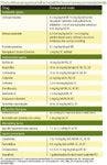

Products for management of bacterial infection or canine parvoviral enteritis
Gram-negative bacterial infections predominate, with E. coli being the most common isolate. However, any bacterial, fungal, parasitic or viral organism may cause sepsis.
Sepsis is diagnosed based on the presence of an underlying infection and identification of systemic inflammatory response syndrome (SIRS). Bacteremia often is not identified and therefore a negative blood culture does not rule out the presence of existing sepsis.
Definitions:
- Systemic Inflammatory Response Syndrome (SIRS): a clinical syndrome caused by systemic inflammation of infectious (i.e., sepsis) or non-infectious origin. In dogs, the diagnosis of SIRS is based on fulfillment of at least two of these criteria: tachycardia, tachypnea, hypothermia or hyperthermia and leukocytosis, leukopenia or greater than 3 percent band neutrophils.
- Sepsis: the systemic inflammatory response to infection
- Severe sepsis: the systemic inflammatory response to infection associated with organ dysfunction and manifestations of hypoperfusion or hypotension
- Septic shock: the systemic inflammatory response to infection with hypotension refractory to volume expansion
- Bacteremia: the presence of bacteria in the blood stream
Pathophysiology
The sequence of events leading to sepsis is complex and not completely understood. In the initial phases of infection, microbial products (e.g., endo-toxin from gram-negative bacteria; exotoxins, peptidoglycans and super antigens from gram-positive bacteria; and fungal cell-wall material) induce systemic inflammation through activation of immune cells, resulting in an imbalance between pro-inflammatory mediators (e.g., TNF) and anti-inflammatory mediators (e.g., IL-10).
Tumor necrosis factor (TNF), IL-1 beta, IL-6, IL-8 and leukotrienes are examples of important pro-inflammatory mediators contributing to the pathologic effects of sepsis in dogs. Ultimately, induction of pro-inflammatory mediators leads to inflammatory cell infiltration, altered thermoregulation, vasodilation, vascular leakage and coagulation.
Clinical effects
Dogs can have either a hyper-dynamic or hypodynamic response during sepsis. The hyperdynamic response is characterized by fever, brick-red mucous membranes, tachycardia and bounding pulses. With disease-process progression, a hypodynamic response characterized by hypotension, pale mucous membranes and hypothermia can be observed.
Often dogs will have GI or respiratory signs associated with the sepsis. Possible serum chemistry profile abnormalities may include hyperglycemia or hypoglycemia, hypoalbuminemia, azotemia, hyperbilirubinemia and elevated serum ALT and/or ALP.
Coagulation abnormalities are common. Anticoagulant proteins (protein C and antithrombin) are significantly decreased and PT, PTT and D-dimer concentrations are significantly increased in naturally occurring sepsis in dogs.
Altered microcirculation and tissue hypoxia may lead to metabolic acidosis in septic dogs. Little is known about the prevalence of organ dysfunction or failure during canine sepsis, although hemodynamic derangement, renal failure and respiratory failure are recognized.
Treatment
The most important aspect of treating sepsis in dogs centers on the identification and eradication of the inciting cause (see "Products for management of canine parvoviral enteritis or bacterial infection" below).
An effort should be made to identify the causative microorganism through cytologic examination and culture. Although stringent effort should be made to identify the cause of sepsis, antimicrobial treatment should not be withheld pending these results.
The use of appropriate broad-spectrum antimicrobial agents is recommended. Since bacteria are the leading cause of sepsis in dogs, typically broad-spectrum antibiotic therapy (e.g., fluoroquinolone plus penicillin derivative) is instituted. The remainder of medical therapy centers on maintenance of tissue perfusion and aggressive supportive care. Treatment should, however, be tailored to the needs of the individual dog.
Cardiovascular support is an important aspect of maintaining good tissue perfusion. Intravenous fluid therapy should be instituted in any dog suspected of having sepsis. For some dogs, the administration of colloids and vasopressors will be indicated. Dogs should be volume-resuscitated prior to the institution of vasopressor therapy because vasopressors may alter microvascular circulation, leading to increased tissue hypoxia.
In one experimental study of canine sepsis, epinephrine was found to adversely affect organ function, systemic perfusion and survival as compared to the use of norepinephrine or vasopressin. Along with maintaining good tissue perfusion, maximizing cellular oxygenation is an important aspect of the treatment of sepsis. This may be accomplished by ensuring good oxygen-carrying capacity, providing supplemental oxygen when indicated and preventing microthrombi formation.
Additional supportive care, including nutritional management, frequent reassessment and good nursing care are key aspects in the management of sepsis. Because bacterial translocation from the GI tract is a substantial concern, the early introduction of preferably enteral or, in dogs with vomiting, parenteral nutrition is paramount. Often these dogs require a feeding tube, and this measure should be taken as soon as possible in a hyporexic or anorexic dog.
Additionally, medication aimed at maintaining normal GI-protective mechanisms (e.g., omeprazole, famotidine, Carafate) should be considered. Careful monitoring for any change in the dog's status (e.g., hypotension, dehydration, organ dysfunction, serum chemistry profile or coagulation abnormalities) is prudent and appropriate therapy should be administered as indicated.
Finally, care should be taken to ensure adequate nursing care, including careful use of aseptic technique during catheter manipulation and frequent turning/movement of the afflicted dog.
Novel therapies
Polymyxin B is a cyclic cationic polypeptide antibiotic that binds the endotoxin that is released from gram-negative bacteria during sepsis, causing activation of inflammatory cells. Endotoxin that is bound to polymyxin is unable to activate inflammatory cells, preventing cytokine production and development of systemic inflammation.
Administration of polymyxin may be an efficacious, affordable and safe means of inhibiting gram-negative, bacteria-induced inflammation dogs. In a placebo-controlled clinical trial, dogs treated with polymyxin (12,500 IU/kg, IM BID) had significantly improved hydration, capillary refill time, pulse quality and significantly lower plasma TNF concentrations than the control group during naturally occurring gram-negative sepsis.
Potential dose-dependent side effects of polymyxin B include respiratory depression, nephrotoxicosis, cardiovascular compromise and neuro-toxicosis. However, the dose needed to achieve anti-endotoxic effects is low, making these side effects unlikely. Polymyxin B is routinely used in the treatment of gram-negative sepsis in horses with good success. Therefore, polymyxin B may be considered as a potential treatment for gram-negative sepsis in dogs.
Activated protein C is an anticoagulant protein. It possesses anti-inflammatory, profibrinolytic and anti-apoptotic effects. During sepsis, there is a clear interrelation between the coagulation system and the inflammatory system. In a multi-center, randomized, controlled, clinical trial, recombinant human-activated protein C decreased mortality from severe sepsis in humans. A canine form of activated protein C is not available but, if developed, could be a promising novel treatment for sepsis in dogs.
Corticosteroids: The use of cortico-steroids for treating sepsis has fallen out of favor due to their immunosuppressive, ulcerogenic and prothrombotic nature. Additionally, clinical trials of the use of corticosteroids in human sepsis have been disappointing. One exception may be the use of low or physiologic doses of corticosteroids for management of relative adrenal insufficiency during sepsis.
In human medicine, relative dysfunction of the adrenal glands does occur during severe sepsis. Corticosteroids have been shown to decrease the need for vasopressors during septic shock in patients with relative adrenal insufficiency. Reports of the effects of corticosteroids on mortality in this situation have been mixed, but lower doses appear to be associated with better outcome. At this time, there is little information about the incidence of relative adrenal insufficiency in dogs with sepsis. Therefore, it is unknown if corticosteroids may be an appropriate adjunctive therapy in dogs. Care should be taken to avoid corticosteroids except in rare cases where adrenal insufficiency is highly suspected and/or documented.
Insulin: Alterations in glucose metabolism during sepsis are well documented. The administration of regular insulin to maintain tight glycemic control has decreased mortality by approximately 40 percent in humans with post-surgical sepsis. Additionally, in experimental models, administration of insulin decreases inflammation and mortality from sepsis. Although this management strategy has not been evaluated in dogs, it warrants further attention and good glycemic control should be a consideration.
Anesthetic/analgesic drugs: Most anesthetic/analgesic drugs possess anti-inflammatory properties during sepsis. Drugs like the opiate sufentanil and the NMDA-receptor antagonist (e.g., ketamine) have been shown to decrease the production of TNF during experimental endotoxemia in dogs. The clinical impact of these findings is unknown and further study is indicated to determine the role of anesthetic/analgesic drugs for the management of sepsis.
Note: Owners do not want their beloved sick dog to be in pain.
Canine parvoviral enteritis or bacterial infection History and physical examination
- Acute gastrointestinal problems in puppies younger than 6 months
- Gastrointestinal signs such as vomiting, diarrhea, anorexia, depression
Laboratory confirmation
- Fecal parvovirus antigen test
- CBC
- Serum chemistry profile and electrolytes
- Intestinal parasites are likely to be present such as hookworms, roundworms, giardia.
Medical and/or surgical procedures
Aseptically place an intravenous or intraosseous in-dwelling catheter.
Provide adequate fluids for reperfusion of vital organs, using lactated Ringer's solution or Normosol-R at a volume and rate adequate to restore perfusion to the vital organ at a supranormal level.
If perfusion is poor, rapidly infuse an intravenous bolus of hetastarch or dextran 70 at a rate of 20 ml per kg for initial resuscitation and provide supplemental oxygen by nasal catheter.
Do not use hypertonic saline solution in this resuscitative process, because the animal is usually severely dehydrated.
Rehydrate with lactated Ringer's solution or Normosol-R at a rate of 3 ml to 10 ml per kg per hour initially until hydration is restored over four hours; maintenance rate is 2 ml to 3 ml per kg per hour.
Note: Using hetastarch or dextran 70, less fluid is lost into the gastro-intestinal tract, and the total volume of fluid required for rehydration is approximately 50 percent of what is used when lactated Ringer's solution or Normosol-R is used alone.
Administer intravenous antimicrobial agents such as first-generation cephalosporins.
If the animal appears to be septic, consider cephalosporins, an aminoglycoside and metronidazole once perfusion has improved.
Palpate the animal's abdomen at least every four hours to detect intussusception.
Give nothing orally until vomiting is controlled.
Flush indwelling catheter with heparinized saline solution every six hours.
Warm or cool the animal as deemed necessary once perfusion has been restored.
Listen for bowel sounds; if decreased or no bowel sounds are detected, put the animal on metoclopramide via the intravenous drip.
Control significant vomiting with administration of metoclopramide or chlorpromazine.
If vomiting is persistent, place a nasogastric tube and suction the gastric contents every one to two hours initially; back off the frequency of suctioning as directed by withdrawal of gastric fluid.
Use the nasogastric tube to give microenteral nutrition.
Once the vomiting has been controlled, begin the administration of oral electrolyte solution supplemented with glucose; this can be done by giving 2 ml to 10 ml of a oral electrolyte solution by dosing syringe, or the oral electrolyte solution can be placed in a fluid bag and dripped continuously into the nasogastric tube at a rate of 2 ml to 10 ml per hour.
Once the animal tolerates the oral electrolyte solution for at least four to six hours, begin liquid nutritional supplementation.
Check monitoring
Packed-cell volume, total plasma solids, blood urea nitrogen, glucose, sodium, and potassium every four to six hours.
Supplement and adjust fluid rate as deemed necessary.
Check perfusion parameters (mucous-membrane color, pulse rate and intensity, capillary refill time, blood pressure, central venous pressure) every two to four hours, and resuscitate with fluids plus or minus hetastarch or dextran 70 infusion as necessary.
Estimate quantity of vomiting, diarrhea and urine output, and record observations every two hours.
Monitor rectal temperature every four to six hours.
Maintenance
Anticipate the problems of poor perfusion, severe dehydration, hypokalemia, hypoglycemia, hypoproteinemia, aspiration pneumonia, sepsis/septic shock, intussusception, hyperthermia or hypothermia, and massive fluid replacement requirements.
Maintain the albumin concentration above 2 g/dl, which likely needs to be done with fresh-frozen plasma on hospital days 2 to 4.
Administer hetastarch or dextran 70 at a rate of 10 ml to 20 ml per kg over four hours, decreasing lactated Ringer's solution or Normosol-R during this time interval, on hospital days 2 and 3.
Owner/client information to discuss
- Cost estimate
- Vaccinate other dogs
- Good sanitation, such as use disinfectants for environmental cleanup
Notes to remember
- Very poor prognosis for Rottweilers
- Subcutaneous fluids may cause sterile abscesses and slough the skin because of poor circulation.
- Update other vaccinations after clinical recovery.
Dr. Hoskins is owner of Docu-Tech Services. He is a diplomate of the American College of Veterinary Internal Medicine with specialities in small animal pediatrics. He can be reached at (225) 955-3252, fax: (214) 242-2200 or e-mail: jdhoskins@mindspring.com.
Newsletter
From exam room tips to practice management insights, get trusted veterinary news delivered straight to your inbox—subscribe to dvm360.


