Study supports the use of AI imaging for canine dermal and subcutaneous masses


An artificial intelligence-based thermal imaging system screens to rule out malignancy
Researchers with HT Vista have published a study1 demonstrating the use of thermal imaging and artificial intelligence (AI) for accurate screening with canine dermal and subcutaneous masses.2 HT Vista is the UK subsidiary of HT Vet, which is a company aiming to increase early detection of skin cancer in dogs. This device from HT Vista would determine whether a mass is benign or needs further investigation of malignancy. Unlike traditional imaging methods, HT Vista assesses disparities in heat transfer rates between masses and adjacent healthy tissues, enhancing the precision of analysis.2

")
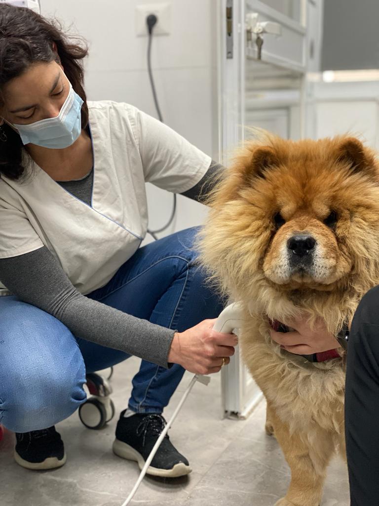
Tali Buber, DVM, veterinary medical director at HT Vet, scanning a dog with new AI imaging device. (Photo courtesy of HT Vista)
The study evaluated over 660 masses between 2020 and 2022 across 2 phases; the first to train the algorithm and the second to validate it.2 Both the training phase and validation phase used client-owned dogs. During the scan, each mass and its adjacent healthy tissue was heated by a high-power Light-Emitting Diode. The device recorded and analyzed the temperature of the tissue and employed a supervised machine learning algorithm to assess whether the mass warranted further examination. The initial study was conducted to gather data for training the algorithm, while the second study verified the algorithm's accuracy by comparing its real-time predictions to the results of cytology and/or biopsy tests.1
Liron Levy-Hirsch, managing director of HT Vista’s UK subsidiary said in the release, “We are thrilled to have scientific research validating the success of the HT Vista device. The veterinary teams who have already adopted the device into their practice are having great success with it, and with the backing of this newly published paper we hope to reach more practices and ultimately save more dog’s lives.”
“It is exciting to see the algorithm improve with every scan. We are very pleased with the results we have and continue to look to the future where we are sure the sensitivity will increase further,” he continued.2
The study results found that the device correctly classified 45 out of 53 malignant masses and 253 out of 378 benign masses (sensitivity = 85% and specificity = 67%). The negative predictive value of the system (percent of benign masses identified as benign) was 97%.1 Researchers concluded that the findings affirm the notion that this innovative system could be used as a decision-support tool, enabling clinicians to differentiate between benign lesions and those requiring further diagnostics, aiding in the rise of early cancer detection. Enhancing diagnostic capabilities holds the potential to advance early treatment. This could make a significant impact since In dogs, cancer accounts for approximately 20% of deaths at 5 years and increases to 40% to 50% from 10 to 16 years of age.3
Tali Buber, DVM, veterinary medical director at HT Vet, said, “I'm excited to be a significant part of bringing innovation to the veterinary profession. The HT Vista development has been extremely satisfying—scanning the cases, comparing the results to the lab reports, enriching the database and working hand in hand with the AI team to improve the algorithm.”2
References
- Dank G, Buber T, Rice A, et al. Training and validation of a novel non-invasive imaging system for ruling out malignancy in canine subcutaneous and cutaneous masses using machine learning in 664 masses. Front Vet Sci. 2023;10. doi.org/10.3389/fvets.2023.1164438
- Newly published paper validates innovative AI-powered screening device for canine dermal and subcutaneous masses. News release. HT Vista. October 2023. Accessed October 30, 2023.
- Bronson RT. Variation in age at death of dogs of different sexes and breeds. Am J Vet Res. 1982;43(11):2057-2059.
Newsletter
From exam room tips to practice management insights, get trusted veterinary news delivered straight to your inbox—subscribe to dvm360.


: An Innovative, Once Daily Canine Treatment That Targets Itch")
Zenrelia™ (ilunocitinib tablets): An Innovative, Once Daily Canine Treatment That Targets Itch
June 25th 2025Finding an effective, safe medication to control itch is essential to maximize patient comfort. Because Zenrelia does not require a loading dose and is administered once daily, Zenrelia may help improve owner compliance and can be cost effective* for pet owners
Read More


